Screw fixation
1. Principles
Anatomic reduction
Perfect anatomic reconstruction is essential for articular fractures to avoid the development of arthritic changes.
Stabilization
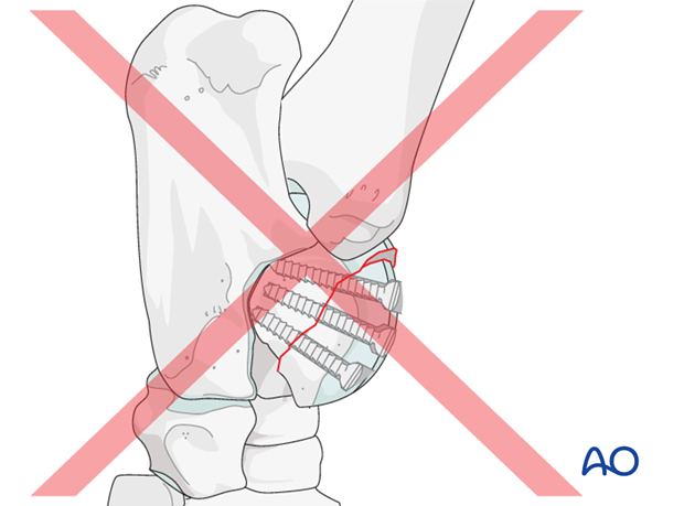
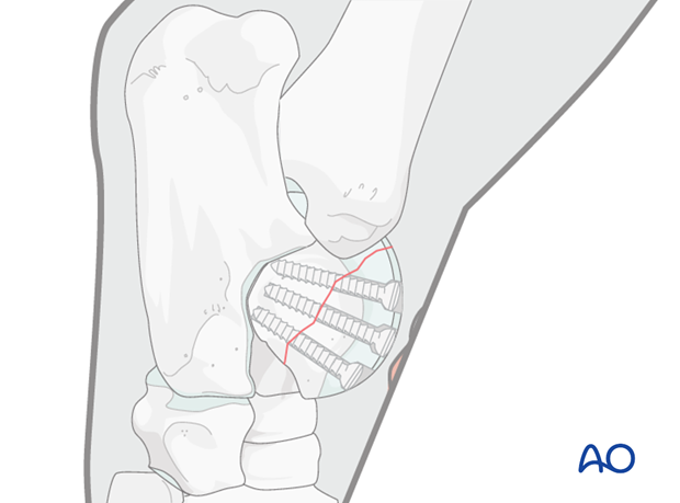
In most cases, two or three 3.5 or 4.5 mm cortex screws are sufficient to stabilize the fracture.
2. Preparation and approach
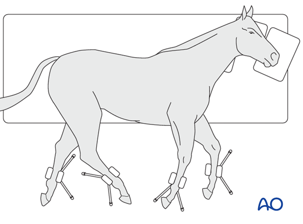
This procedure is performed with the patient placed in lateral recumbency and through stab incisions.
3. Arthroscopic evaluation
The arthroscope is introduced through a medial approach.
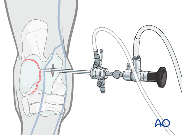
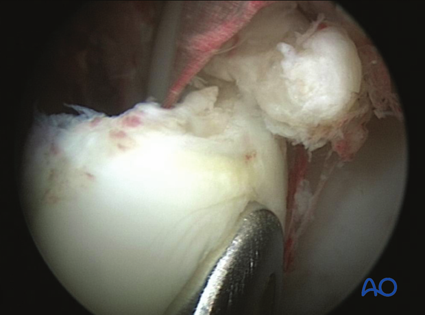
After arthroscopic evaluation of the fracture, the joint is flushed and the blood and small fragments removed.
4. Reduction
Most trochlear fractures are only slightly dislocated and reduction can be easily achieved by compression with blunt instruments.
Anatomic reduction is confirmed by intraoperative radiographs as well as arthroscopic evaluation.
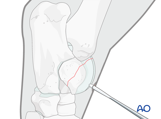
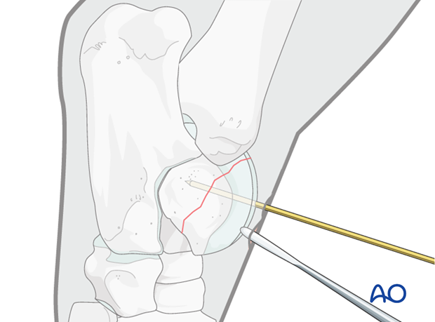
When perfect reduction is achieved, it is temporarily held in place by a temporary interfragmentary K-wire.
5. Screw fixation
Preparation of the drill hole
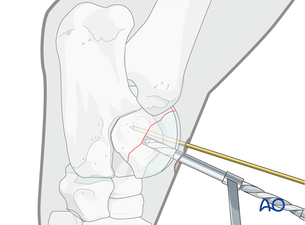
A stab incision is made over the planned screw site followed by a 4.5 mm glide hole drilled perpendicular to the fracture plane from the dorsal aspect of the ridge. Penetration of the drill bit is continuously monitored radiographically or fluoroscopically.
Care is taken to avoid undue penetration of the parent portion of the bone with the glide hole.
Preparation of the thread hole
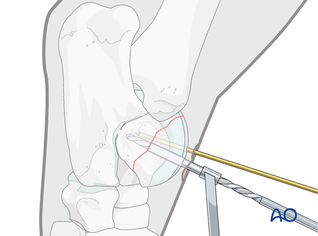
The 3.2 mm drill guide is inserted into the glide hole and the thread hole is prepared.
In most cases one cannot go through the entire talus and has to create a blind thread hole.
Countersinking
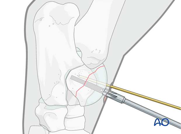
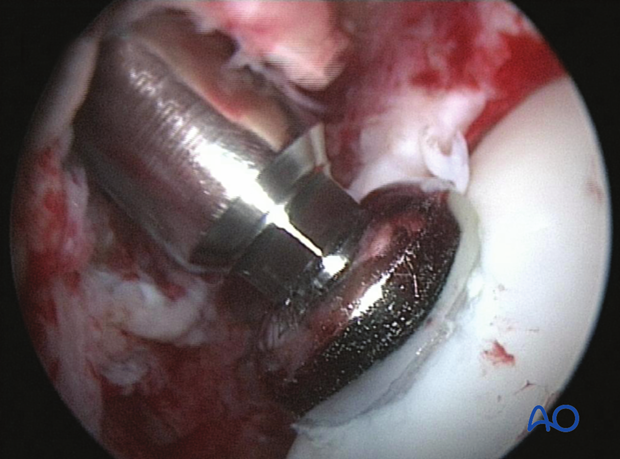
A deep countersink depression is prepared in the articular surface to accept the entire screw head.
Measuring screw length
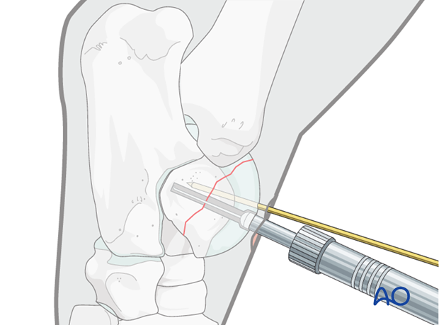
A depth gauge is introduced to measure the required screw length.
With a blind hole, the screw should be 3-4 mm shorter than the measured depth.
Screw insertion
The hole is carefully tapped.
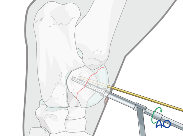
The first screw is inserted and thoroughly tightened.
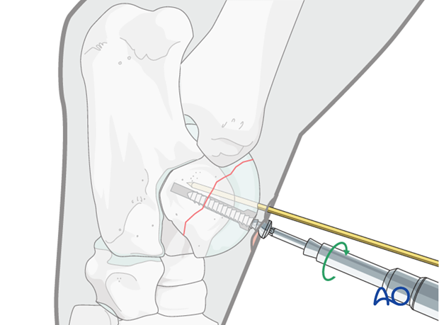
One or two additional screws are inserted applying identical technique.
The arthroscopy portal and the stab incisions prepared for screw insertion are closed by simple interrupted skin sutures.
Radiographic confirmation of the final construct
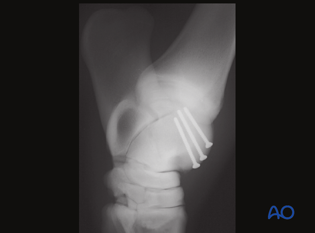
6. Case example
Case example of a 16 year old Thoroughbred.
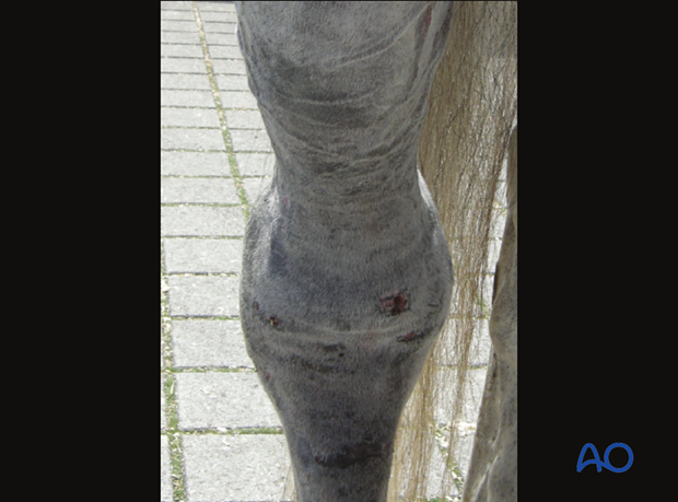
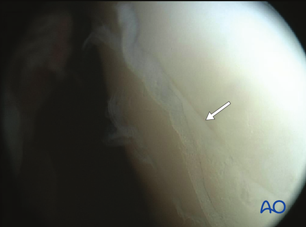
Fracture line as identified under arthroscopic supervision.
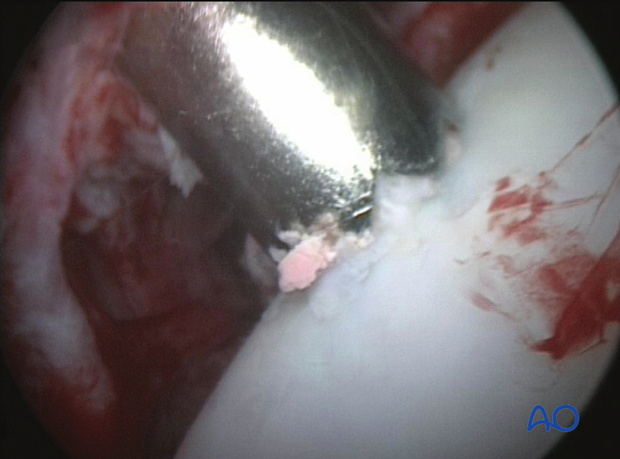
Drilling the glide hole.
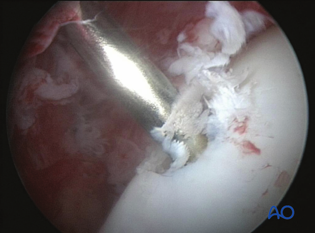
Determining the screw length with the depth gauge.
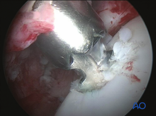
Tapping of the thread hole.
Screw insertion.
7. Aftertreatment
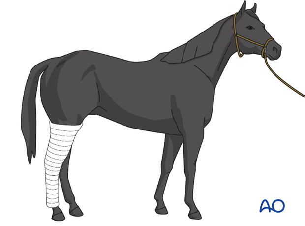
A bandage is applied, the horse recovered, and maintained for two weeks. The bandage should be changed every 5 days.
The sutures are removed after 10 days.
The horse is kept in a stall for 4 weeks. Hand-walking exercises are indicated for another 4 weeks before the horse can be put back into training.
Follow-up x-rays are taken 8 weeks after surgery to evaluate fracture healing.
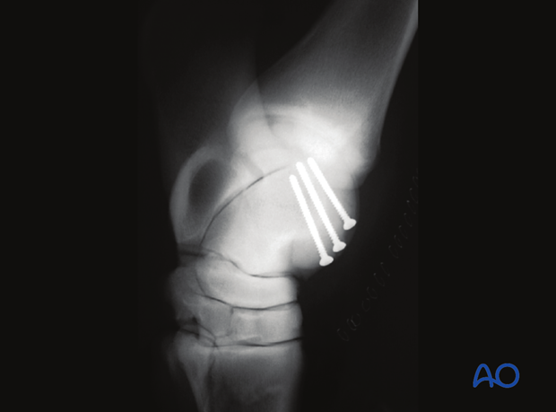
Radiographic control after two years. The fracture has healed completely and no signs of degenerative joint disease can be seen.