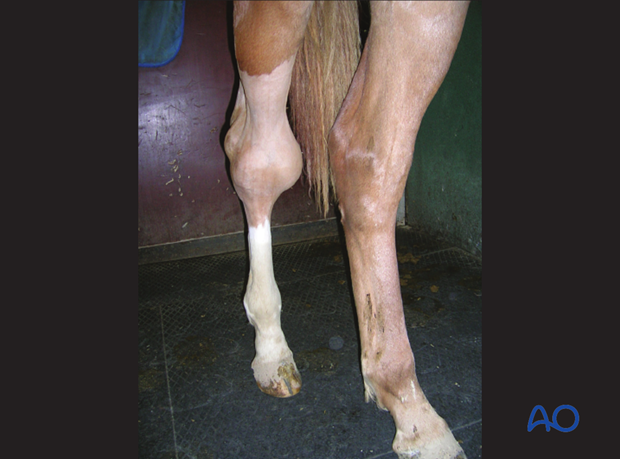
Talus: Sagittal fractures
Fracture characteristics
Sagittal fractures are usually caused by twisting forces and are mostly closed.
They usually cause very severe lameness.
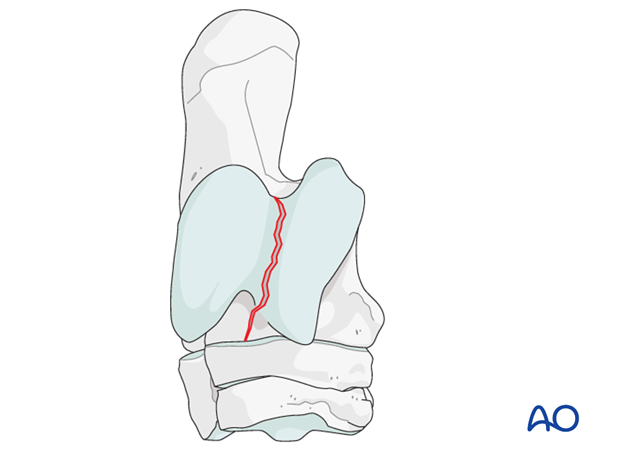
These fractures displace only minimally and are therefore difficult to detect radiographically. Computed tomography is the imaging technology best suited for a detailed diagnosis, which is a prerequisite for proper screw placement.
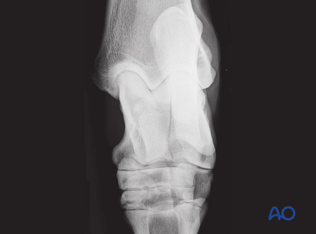
Incomplete sagittal fractures of the talus are rare and according to Davidson et al., the left talus is more commonly affected.
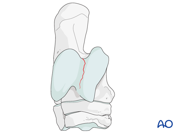
Fractures of the talus
Talar fractures can occur after an external trauma or after twisting or shearing of the tarsus. They vary from simple fragmentation of small portions of the trochlear ridges...,
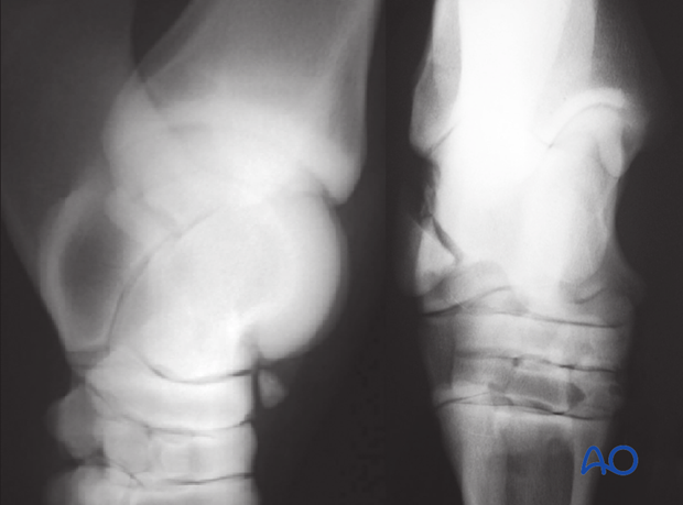
... fractures of the body of the talus...
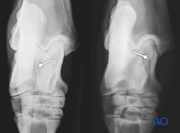
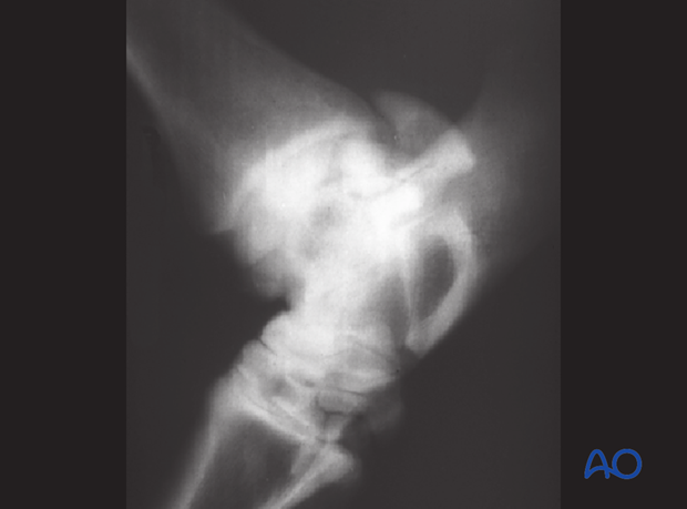
... to completely disruptive multifragment fractures of the entire body of the talus.
Clinical signs and imaging
Clinical signs can vary considerably, but very often horses show an acute severe weight bearing lameness (Grade III to V). Significant effusion of the talocrural joint is seen with most fractures of the talus. Fissures can be difficult to find radiographically. Therefore, several projections with different angles including flexed and skyline projections of the tarsus are necessary.
CT imaging is very useful to detect fissure lines and help to understand the configuration of the fracture.