Screw fixation
1. Principles
Choice of treatment
For the treatment of comminuted fractures, often a combination of several techniques is required, such as arthroscopy and intraoperative radiographic control. Those techniques are described in the complete sagittal fractures and large trochlear ridge fractures.
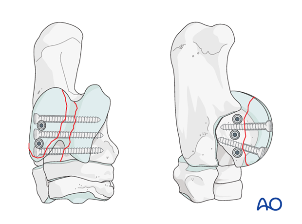
If the comminution is that severe, that sufficient stabilization cannot be achieved, euthanasia has to be discussed.
2. Preparation and approach
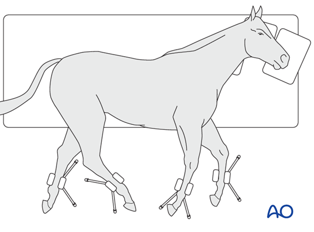
This procedure is performed with the patient placed in lateral recumbency and through stab incisions.
3. Case example
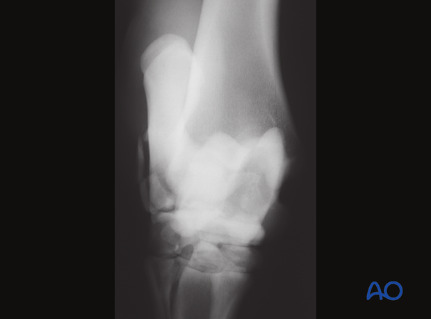
Case example of a 16 year old Thoroughbred with a complete sagittal fracture in conjunction with a medial trochlear ridge fracture of the talus. The fracture is repaired by means of several screws inserted in lag fashion to fix both fracture planes.
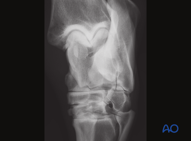
The first screw is inserted to provide interfragmentary compression across the medial trochlear ridge fracture.
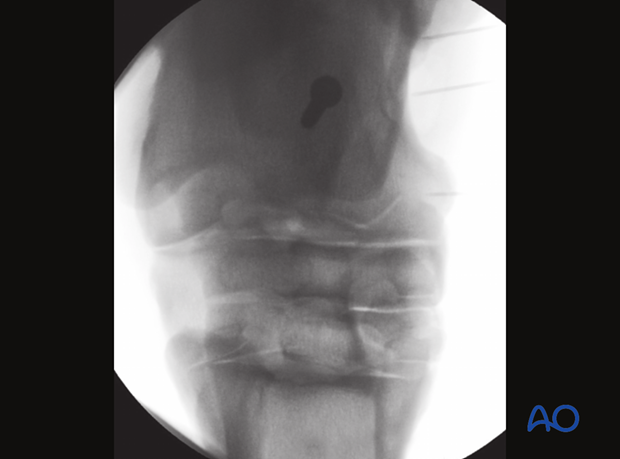
The second screw is inserted to fix the sagittal talus fracture.
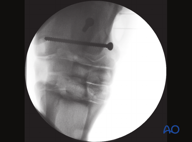
The third screw is inserted again across the sagittal fracture. Intraoperative x-ray reveals that the screw enters the fracture of the medial trochlear ridge fracture and had to be removed.
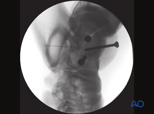
Insertion of the fourth screw provides further compression across the medial trochlear ridge fracture.
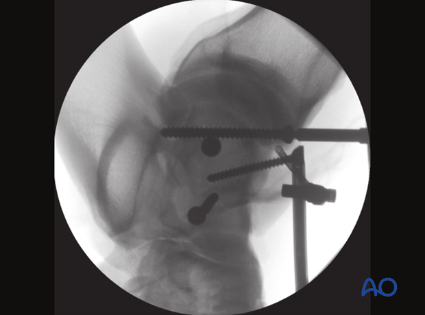
Additional intraoperative imaging revealed that the third screw was partially placed into the fracture line of the trochlear ridge fracture. The screw was therefore replaced.
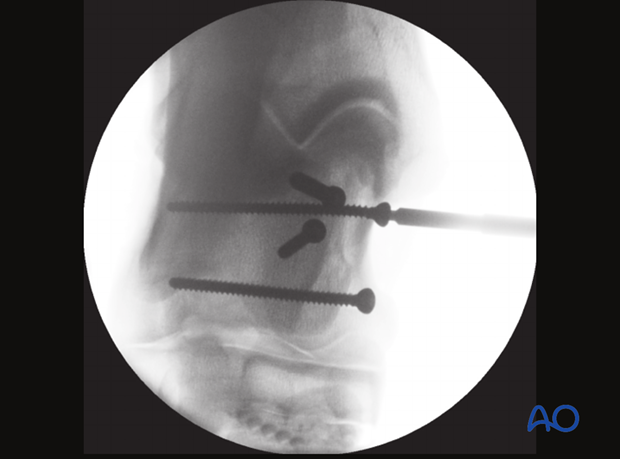
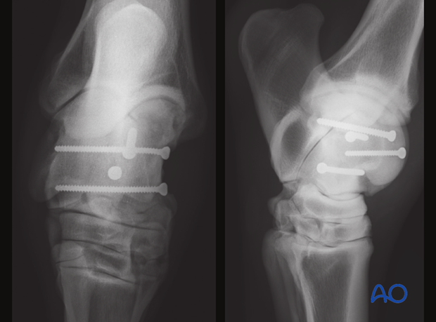
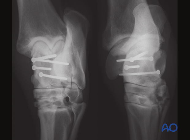
Final radiographic assessment reveals perfect reconstruction of both fracture planes.
4. Aftertreatment
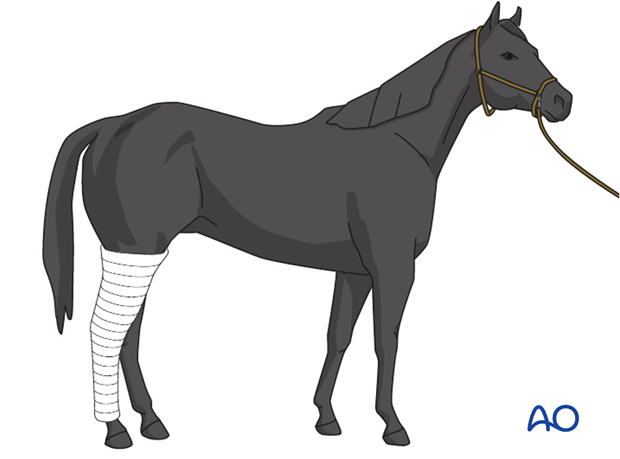
A bandage is applied, the horse recovered, and maintained for two weeks. The bandage should be changed every 5 days.
The sutures are removed after 10 days.
The horse is kept in a stall for 4 weeks. Hand-walking exercises are indicated for another 4 weeks before the horse can be put back into training.
Follow-up x-rays are taken 8 weeks after surgery to evaluate fracture healing.