Screw fixation
1. Preparation and approach
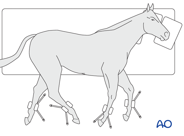
This procedure is performed with the patient placed in lateral recumbency and through stab incisions.
2. Screw fixation
Reduction
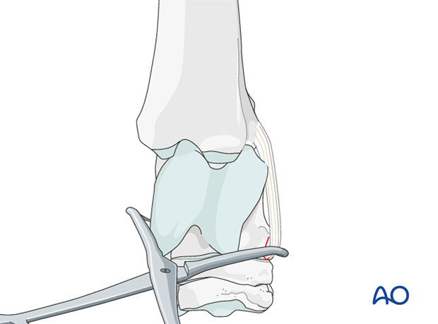
The fracture is reduced with pointed reduction forceps.
Anatomic reduction is confirmed by intraoperative radiographs.
Reduction is maintained with the pointed reduction forceps.
Planning the screw positions
Before insertion of the screw, the optimal screw location is chosen with the help of needles.
One needle is placed into the proximal intertarsal joint and one needle into the talocalcaneal joint (yellow). An additional needle is placed at the planned screw position (blue).
With intraoperative radiographs the proposed position of the screw is confirmed.
Screw insertion
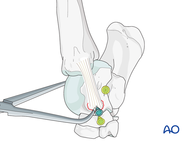
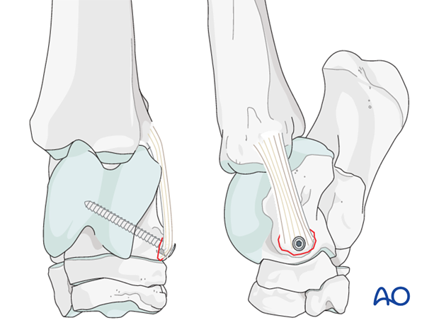
A 4.5 mm cortex screw is introduced in lag fashion.
Pitfall
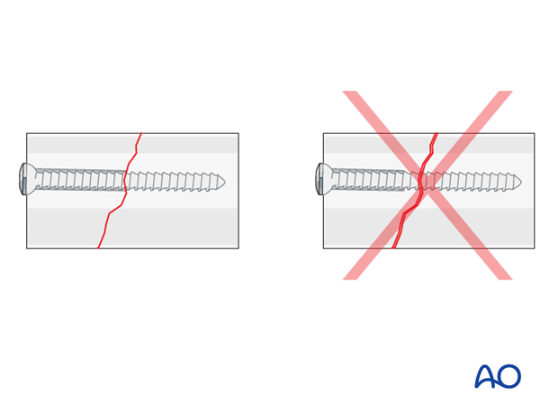
Remember to drill the glide hole long enough, across the fracture line. Too short a drill hole does not allow for interfragmentary compression.
Radiographic example
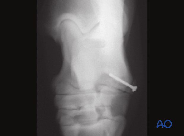
Fixation of a small fragment of the distal lateral trochlear ridge with one 3.5 cortical screw.
3. Resection
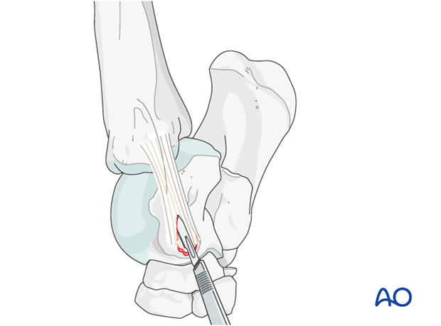
If a small fragment is to be removed, the attachment of the collateral ligament to the fragment needs to be transected, before the fragment can be taken out.
4. Aftertreatment
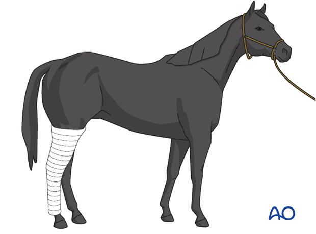
A bandage is applied, the horse recovered, and maintained for two weeks. The bandage should be changed every 5 days.
The sutures are removed after 10 days.
The horse is kept in a stall for 4 weeks. Hand-walking exercises are indicated for another 4 weeks before the horse can be put back into training.
Follow-up x-rays are taken 8 weeks after surgery to evaluate fracture healing.