Imaging
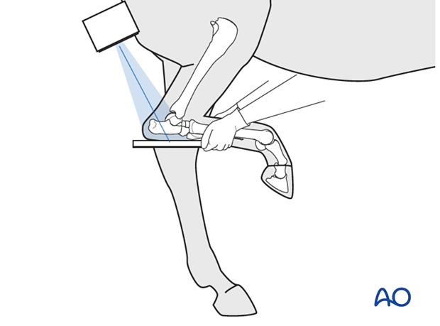
Many tarsal fractures are not extensively displaced and initially may not be evident on routine radiographs. So multiple radiographic projections of the tarsal bones are important and special views like stressed views, skyline projections (see left) are mandatory.
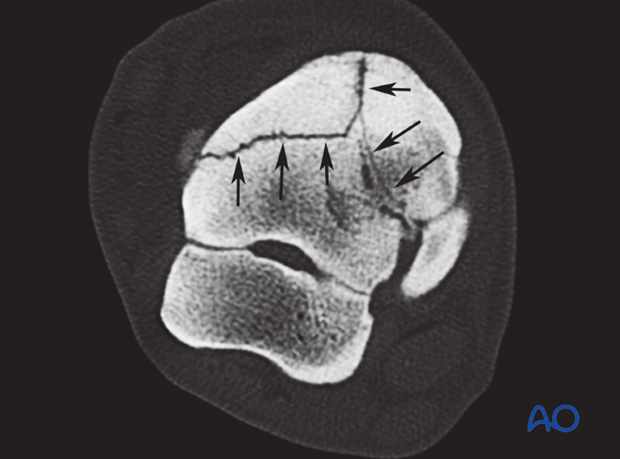
CT and MRT imaging are very useful to find small fractures lines and define their precise configuration. For the correct diagnosis as well as for planning perfect screw position, three dimensional reconstruction of the injured tarsus with CT or MRT studies is often necessary.
Left: Cross-sectional CT image of a comminuted fracture of the central tarsal bone.