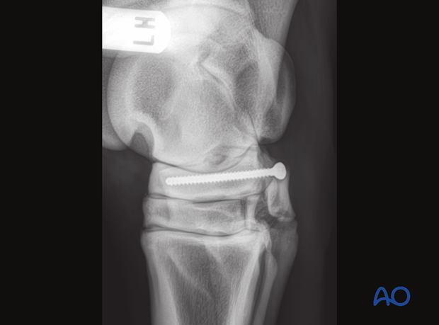
Screw fixation
1. Preparation and approach
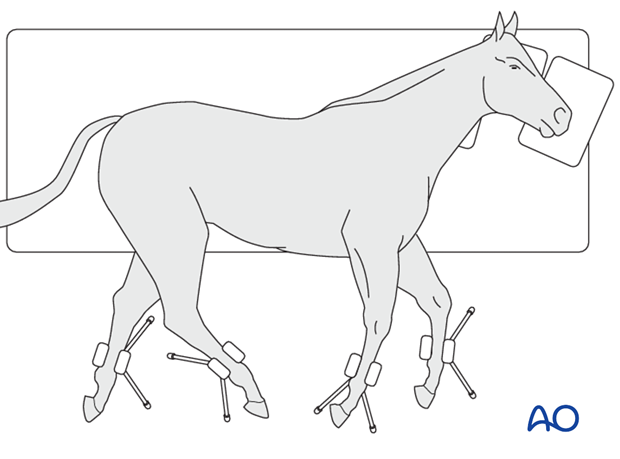
This procedure is performed with the patient placed in lateral recumbency and through stab incisions.
2. Surgical technique
Planning the screw position
The optimal screw position is chosen based on the fracture configuration seen on CT images.
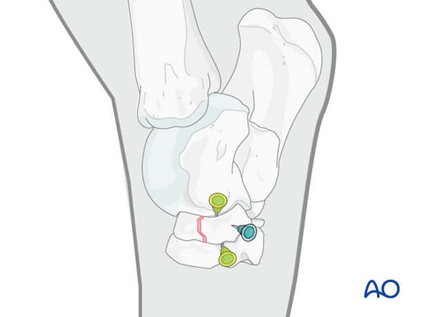
The screw position is determined with the help of needles. One needle is placed into the proximal intertarsal joint, one into the distal intertarsal joint and one at the intended screw position.
Stab incision
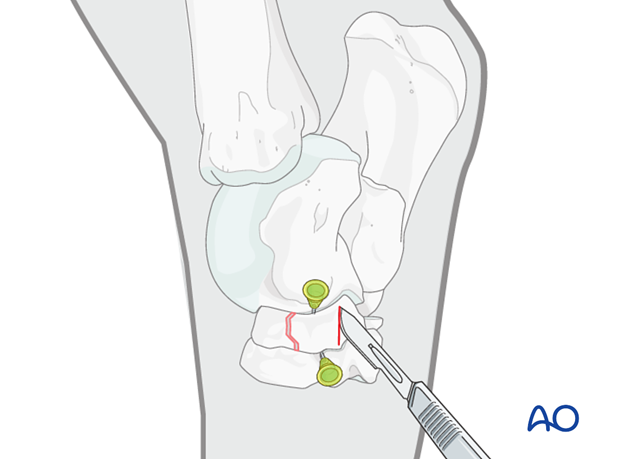
A stab incision is placed over the planned screw position on the medial side of the tarsus over the central tarsal bone.
Drilling the glide hole
A 4.5 mm glide hole is drilled perpendicular to the fracture plane in to the medial aspect of the central tarsal bone.
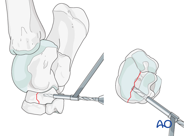
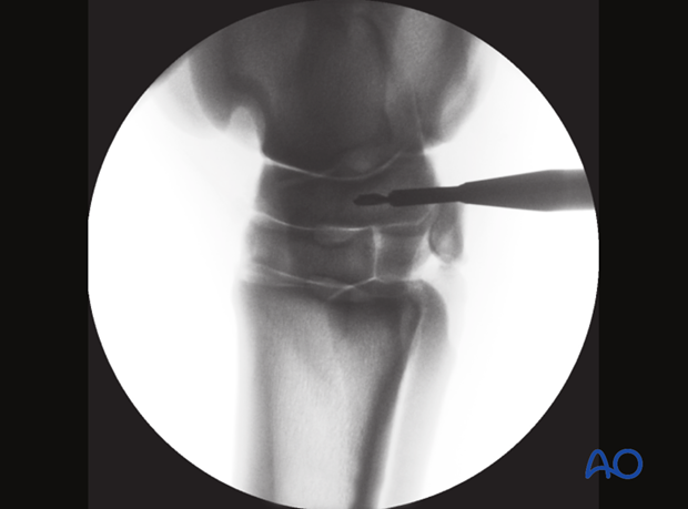
Intraoperative radiography or fluorosocpy aids in recognizing when the glide hole reaches or just passes the fracture plane.
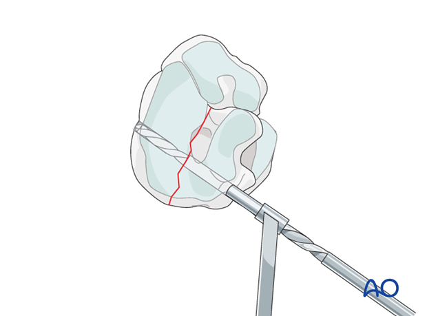
Preparing the thread hole
The 3.2 mm insert drill sleeve is inserted into the glide hole and the thread hole drilled across the rest of the bone.
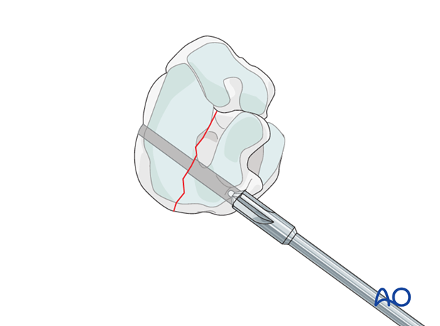
Countersinking
A countersink depression is prepared to accept the screw head.
Measuring screw length
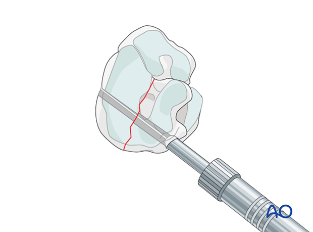
The depth gauge is introduced to determine the required screw length.
Inserting the screw
The hole is tapped and a 4.5 mm cortex screw of predetermined length is inserted and tightened.
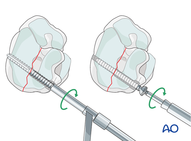
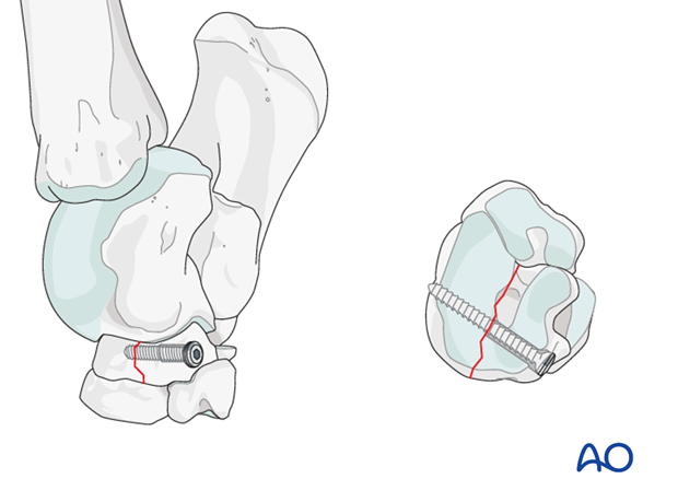
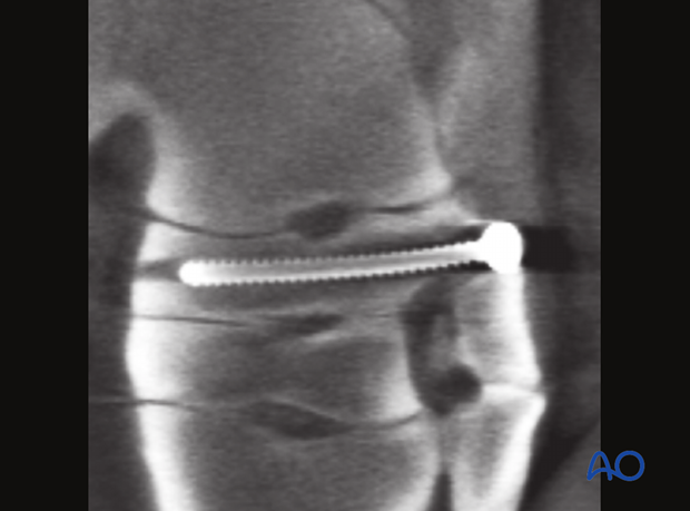
Correct screw placement is confirmed using the C-arm before the stab incision is closed in two layers with interrupted skin sutures.
3. Aftertreatment
Recovery
A support bandage that includes the entire tarsal region is applied and the horse is recovered from anaesthesia with manual assistance.
Aftercare
The limb is bandaged for 15 days. The horse is confined to a box stall for 2 months, after which time it is put on a small paddock or another 2 months. The sutures are removed 10 days postoperatively.
Implant removal
If the lameness persists after surgery, it may be necessary to remove the screw via a medial stab incision. It is believed that postsurgical pain is caused by the rigid implant in a less rigid bone. This is supported by the observation that after implant removal horses have a higher rate of return to athletic activity.