Plate fixation
1. Preparation and approach
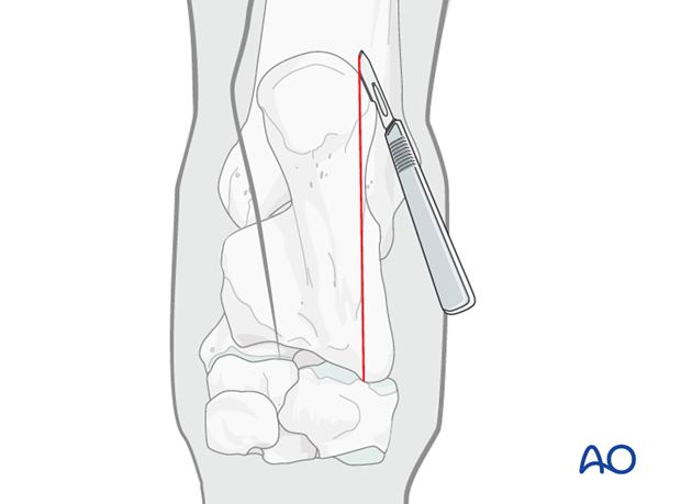
This procedure is performed with the patient placed in lateral recumbency and through the approach to the calcaneal shaft.
2. Reduction and Fixation
Reduction
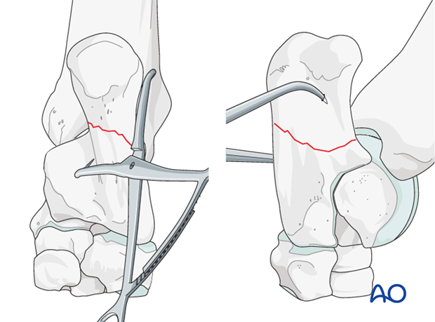
Reduction of the fracture is achieved with a pointed reduction forceps.
Anatomic reduction is essential and should be confirmed with intraoperative radiographs.
Interfragmentary screw
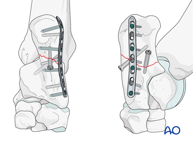
Dependent on the configuration of the fracture plane, either an interfragmentary cortex screw in lag fashion is applied distant from the plate (as shown in the illustration) or through the plate.
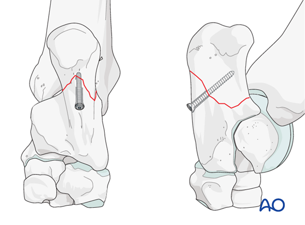
Plate application
A broad 4.5 mm locking compression plate is anatomically contoured to the bone and applied to the plantarolateral surface of the calcaneus. In this case an interfragmentary screw should be placed through the plate (arrow).
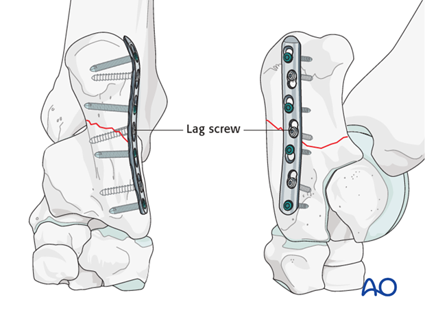
Occasionally, it may not be possible to apply a broad plate. In these cases, a narrow LCP should be applied in conjunction with multiple interfragmentary lag screws or two dynamic compression plates (DCP) should be used.
3. Aftertreatment
Broad spectrum antibiotics are administered for 3 weeks.
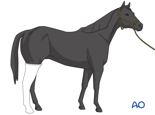
The limb is placed in a full limb cast for three weeks. If necessary it is encouraged to change the cast after 7 to 10 days when the initial swelling has subsided. This is followed by a cast that can be snugly applied to the limb.
The horse is kept in a stall for 4-8 weeks. Hand-walking exercises are indicated for another 4-8 weeks before the horse can be put back into training.
Follow-up radiographs are taken 8 weeks after surgery to evaluate fracture healing and prescribe further postoperative management.
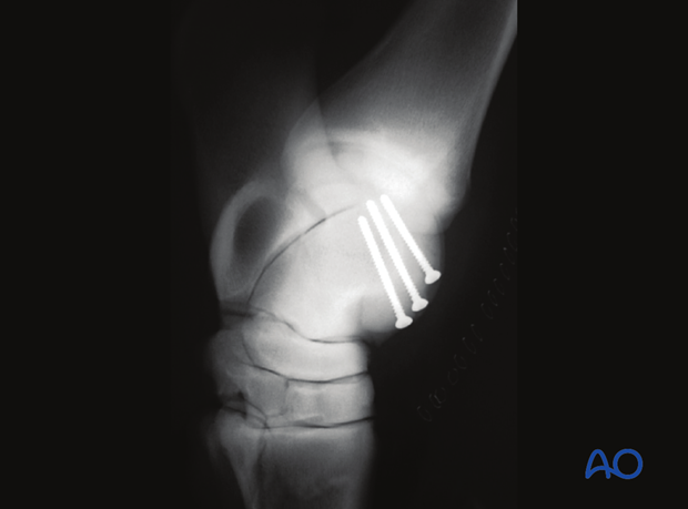
Radiographic control after two years. The fracture has healed completely and no signs of degenerative joint disease can be seen.