Stabilization
1. Principles/General considerations
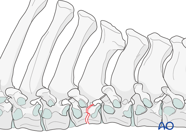
The more common sites of fracture of vertebral bodies include the first three thoracic vertebrae,
around T12 (the area of greatest lateral bending and axial rotation), and the lumbar vertebrae. Dorsal spinous process fractures tend to occur at or near T6.
If the thoracolumbar spinal cord is seriously disrupted, horses should be euthanized immediately. If the spinal cord is intact, stabilization of the vertebrae using pins can be attempted.
There are few reports describing surgical treatment of thoracolumbar fractures in foals.
2. Preparation and approach
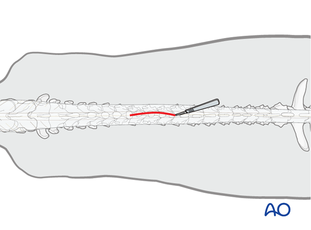
This procedure is performed with the patient positioned in sternal recumbency through the dorsal approach to the thoracolumbar spine.
3. Reduction
Reduction of these fractures is not possible.
4. Stabilization
Stabilization with Steinmann pins and polymethyl methacrylate bone cement can be possible. Sometimes a lumbar dorsal laminectomy can be performed.
Pin insertion
Pins are carefully inserted with powered equipment.
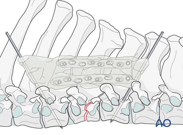
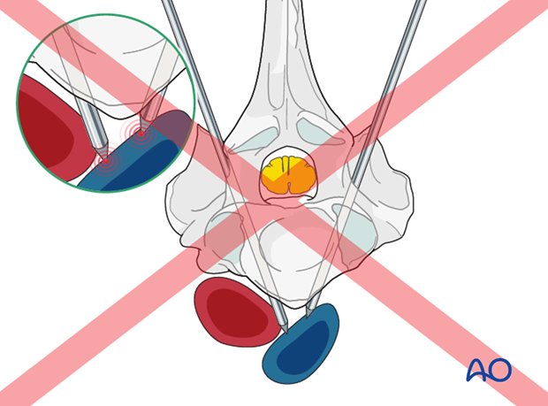
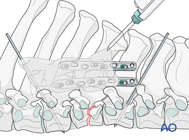
The caudal pin is inserted caudal to the articular processes and lateral to the vertebral canal. They are angled ventrocaudally 30 degrees off the vertical line and towards the midline 20 degrees off the vertical line. The pins should barely exit the ventral portion of the vertebrae.
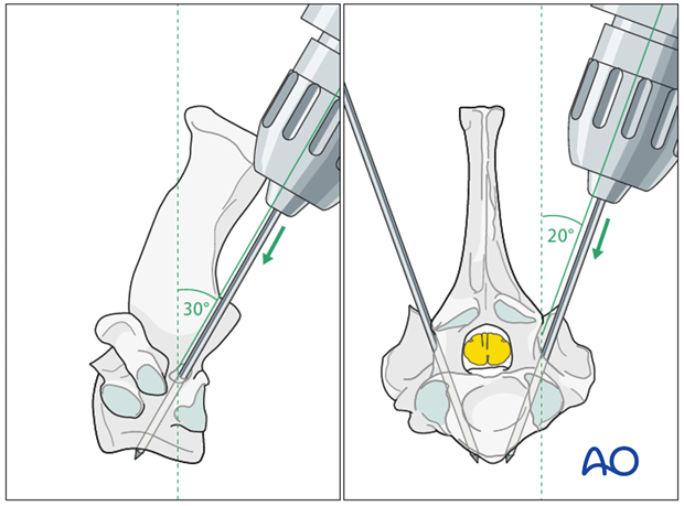
Note: Attention should be paid not to injure the aorta and vena cava, which are running near to the ventral aspect of the vertebrae.
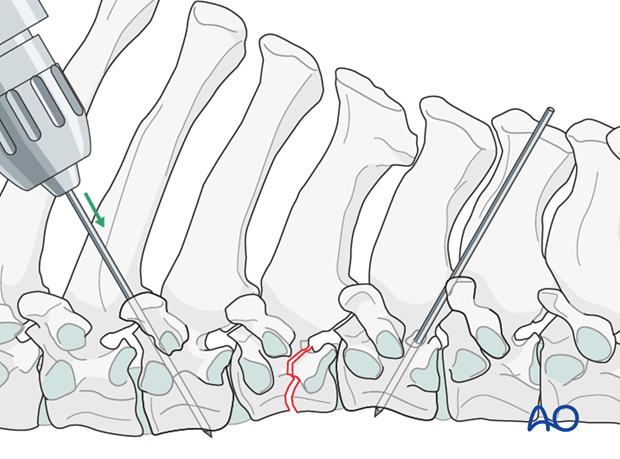
The cranial pins are applied in a similar fashion in a cranioventral converging technique.
With the Steinmann pins in place a pair of plates is applied to the dorsal spinous processes.
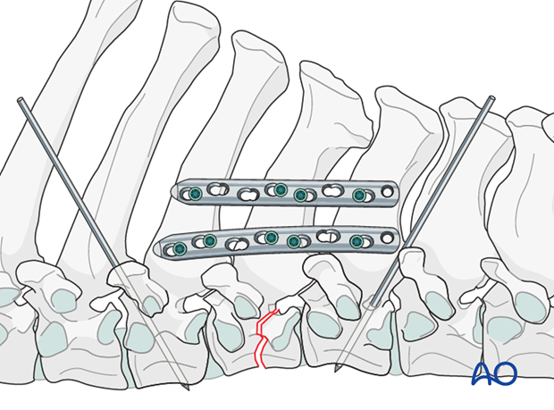
Final rigidity is provided by connecting the Steinmann pins with cold-curing polymethyl methacrylate (PMMA).
PMMA is molded around the plates and the pins.
5. Closure
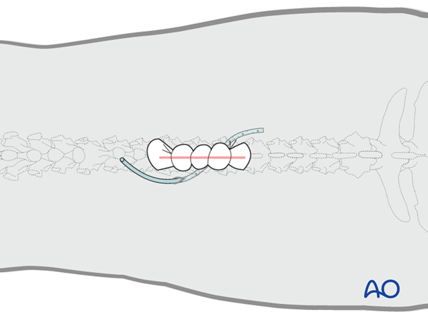
One deep layer and the subcutaneous tissue are closed in simple continuous patterns. The skin is closed with staples. A stent bandage is applied and covered with an adhesive barrier drape to keep the incision clean and dry during recovery.
Suction drains are facultative.
6. Aftercare
Following surgery, antibiotics and NSAIDs are routinely administered for 3 days. If indicated, they need to be continued.
Routinely follow up radiographs are taken immediately after surgery and after 2 and 4 months.
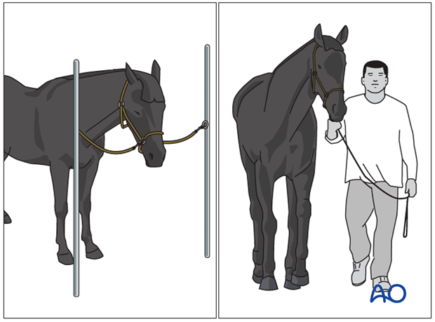
The rehabilitation protocol includes 2 months of stall confinement, followed by 1 month of hand-walking, and 2 months of progressive exercise.
Only when the ataxia has completely disappeared, the horse can return to training or other activities.
Implant removal
There is no need for implant removal, except in cases of implant loosening or surgical site infection.
