Complications in distal femoral fractures
1. General considerations
The most common complications in distal femoral fractures are:
- Malalignment with or without patellar luxation
- Implant failure
- Osteoarthrosis
- Quadriceps contracture
- Nonunion
2. Malalignment with or without patellar luxation
Anatomic alignment is always desirable. Malalignment can occur secondary to:
- Under-reduction of distal femur or physeal fractures, resulting in reduced range of motion and impaired tracking of the patella
- Poor intraoperative alignment of the fracture
- Fixation failure
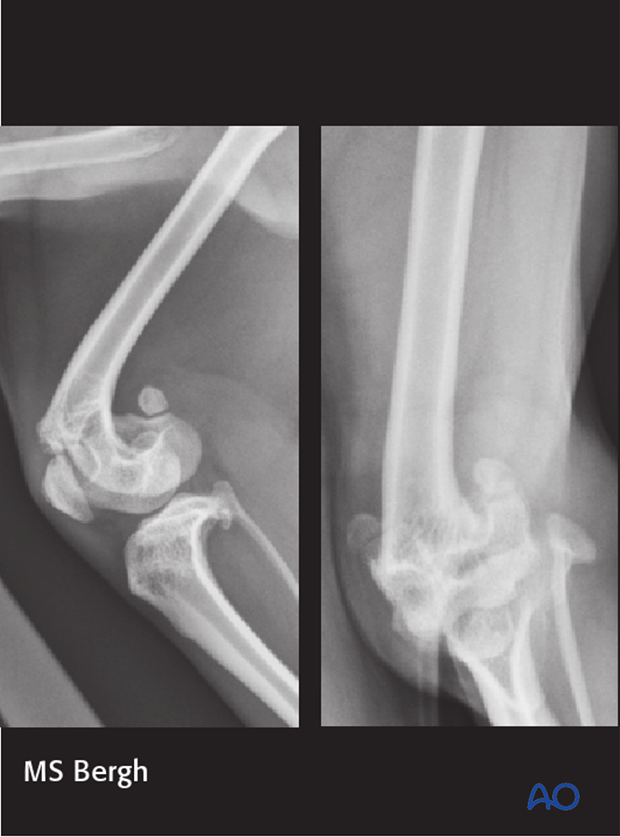
The radiographs show a malunion of a distal femoral fracture in a 3-year-old mix breed that was managed non-surgically. The condyles are under reduced and the metaphyseal segment is impinging on patellar tracking. In addition, there is marked femoral valgus. Function of the limb was poor.
Intraoperative malalignment may occur due to a lack of recognizable landmarks that aid the surgeon in proper alignment of the limb.
Malalignment on the frontal (varus or valgus) or axial plane can severely compromise limb function and result in patellar luxation.
Limb alignment can be assessed by clinical evaluation and intraoperative fluoroscopy or radiographs.
Radiographs show malreduction of the distal physeal fracture resulting in a rotational malalignment and under-reduction of the distal fragment. Note the femoral head anteversion.
3. Implant failure
Implant may fail by bending, breaking or loosening. This is frequently due to inadequate stabilization of the fracture site.
Implant removal or replacement might be needed depending on the stage of healing at which the failure is identified.
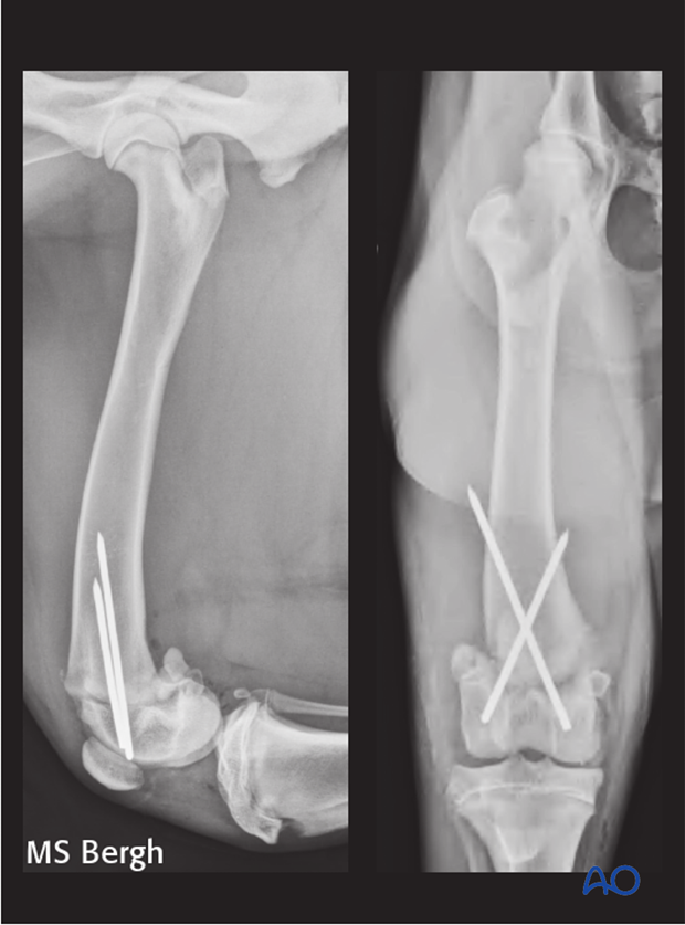
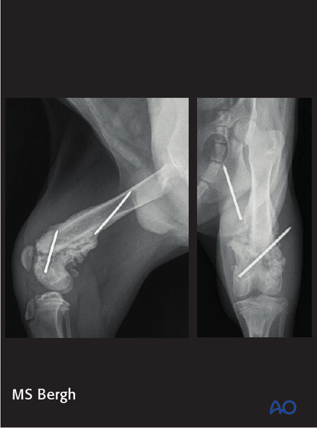
Images show implant loosening and migration. Following repair of a 33-A1 fracture with cross pins. Note loss of reduction of the fracture and significant healing occurring at the time of these radiographs. Despite implant failure, rotational and frontal plane alignment is good.
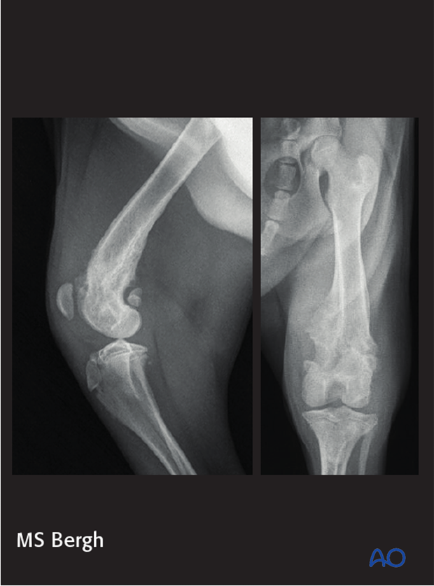
Due to the advanced healing stage the loose implants were removed and an abrasion sulcoplasty was performed in the proximal trochlear groove to improve patellar tracking. These radiographs were taken 4 weeks after implant removal. Notice progressive healing and remodeled distal femur. Clinical function was good.
4. Osteoarthrosis
Osteoarthrosis most commonly occurs following articular fractures. This can be due to the trauma of the original injury, secondary to the surgical trauma or poor reduction of the articular surface.
5. Quadriceps contracture
Quadriceps contracture is a complication seen in young dogs.
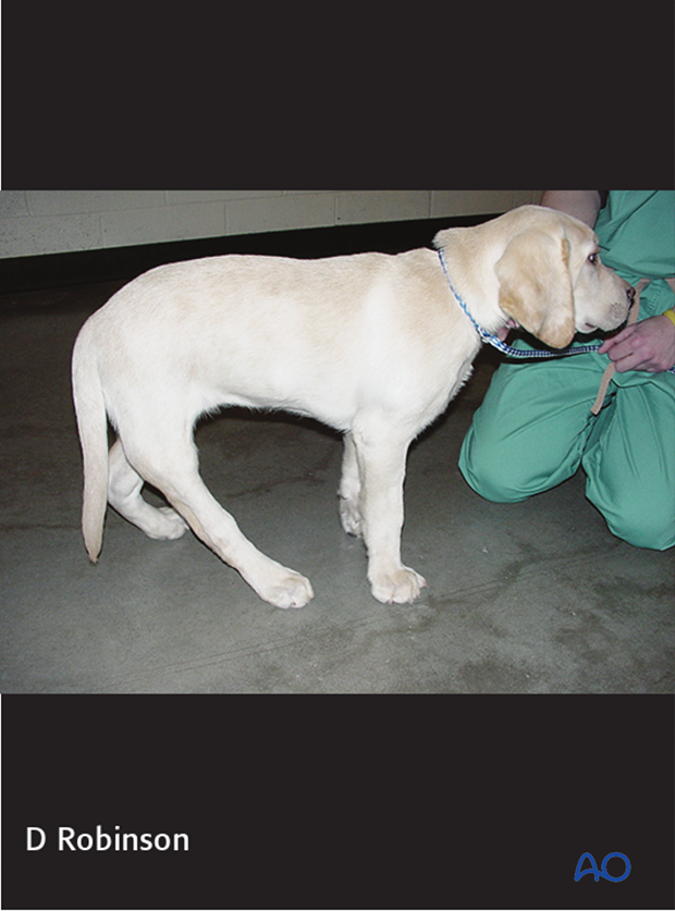
Patients will usually present with severely hyperextended stifle and tarsal joints which are unable to be flexed.
The causes are aggressive surgical approach, poor soft-tissue handling, inadequate fixation and external coaptation such as splinting.
There is no cure for quadriceps contracture and the limb usually has to be amputated to improve mobility and comfort.
Adequate surgical technique, adequate stabilization and early mobilization are key to avoid this complication.
6. Nonunion
Nonunion is the result of a combination of biological and/or mechanical errors that interfere with the bone healing process.
In the distal femur the bone healing potential is usually high and nonunions are result of inadequate stabilization.
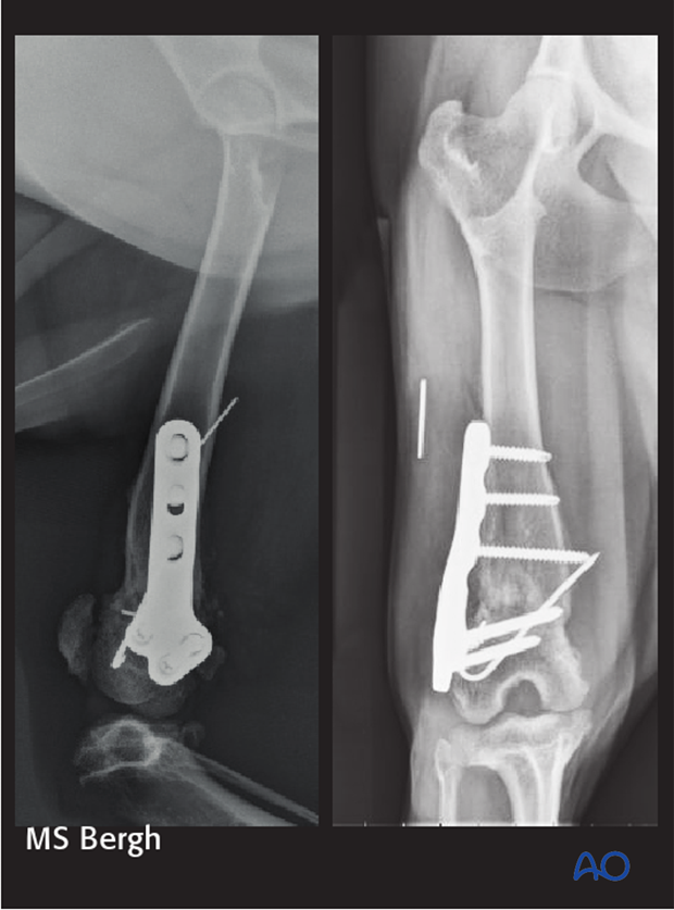
Images show a viable nonunion of a distal femoral fracture repaired with K-wires.
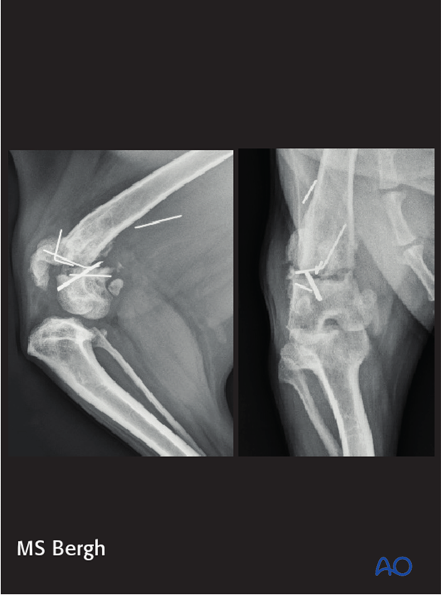
This fracture was revised using a locking compression plate, screws and a K-wire. Autogenous cancellous bone graft was placed at the fracture site.
Postoperative radiographs at 4 weeks show progressive bone union of the fracture. Clinical function was good.
