Percutaneous approach to the dog distal femur
1. Indications
A percutaneous approach is used prior to pin placement when a minimally invasive approach is preferred, and the fracture can be reduced in a closed fashion.
It is most commonly used with 33-A1 and 33-A2 fractures.
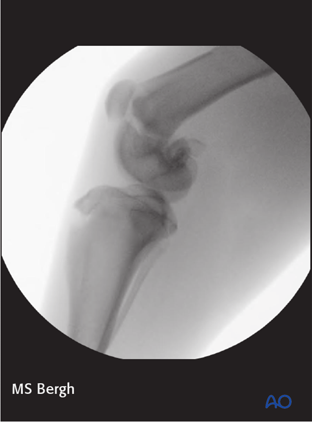
Fluoroscopy or intraoperative radiography is used to ensure appropriate reduction with this approach.

2. Skin incision
A skin incision is made on the regions of the desired pin placements. The subcutaneous fat and superficial fascia are incised directly under the skin incisions.
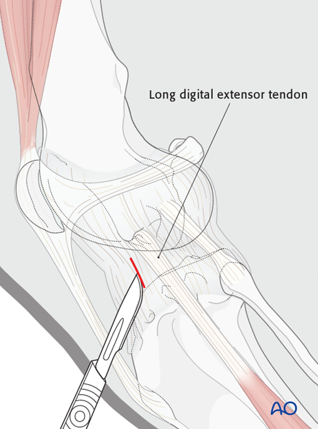
A small incision is made into the joint capsule and the condyle is visualized.
Note: Damage to the articular surface should be avoided. The long digital extensor tendon should be carefully avoided in this approach.

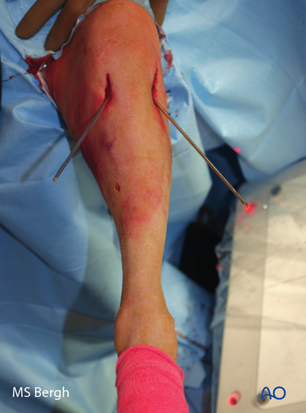
Intraoperative photo of cross pins placed percutaneously.

Closure
Absorbable suture material is used to close the joint capsule and subcutaneous tissue. The skin is routinely closed with nonabsorbable sutures.


