External fixator
1. Principles
General considerations on the external fixator can be found here.
In the tibia, the type 1 fixator should be placed medially. Type 1a, 1b, 2 and 3 frames can be used in the tibia.
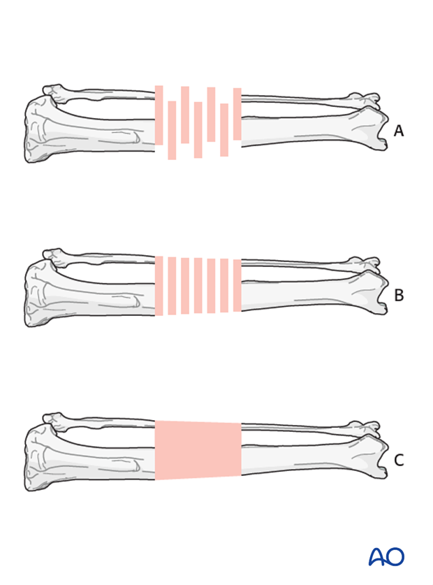
Illustrations of fracture types
B) Comminuted fracture aligned biologically
C) Fracture aligned and biologically stabilized
2. Preparation and approach
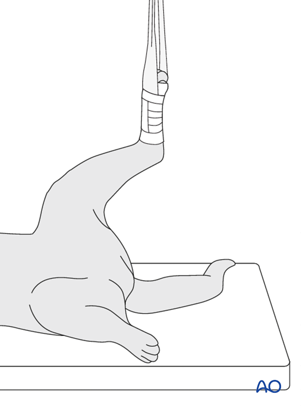
This procedure is performed with the patient in dorsal recumbency.
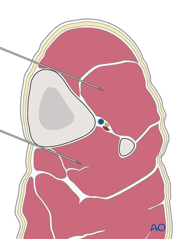
The fixator pins must be placed in the center of the bone. Two hypodermic needles or palpation can be used to locate the bone during closed application of the fixator.
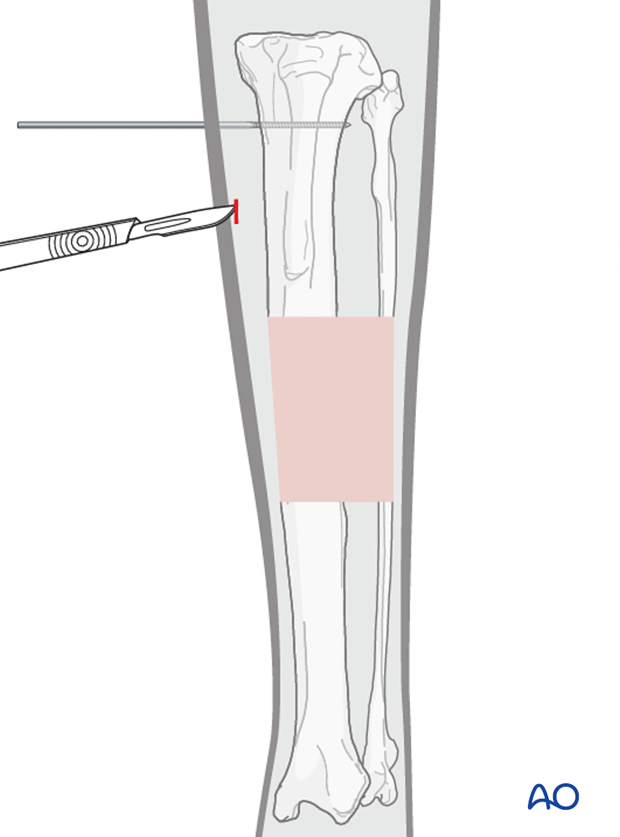
A stab incision is performed at the desired location of pin placement. It is preferred to incise between muscles.
3. Pin placement and insertion
Pin selection
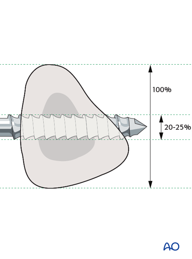
The pin diameter should be 20-25% of the bone diameter. Positive profile pins should be used.
Predrilling
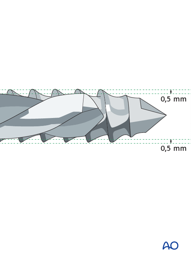
Pre-drilling the hole is strongly recommended prior to pin insertion. The diameter of the drill bit should be 1mm smaller than the pin diameter.
Pin insertion
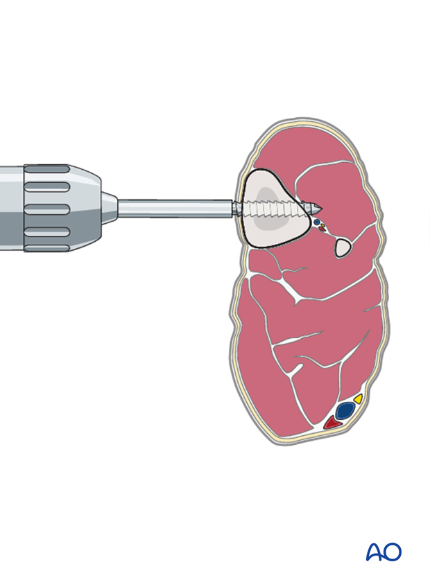
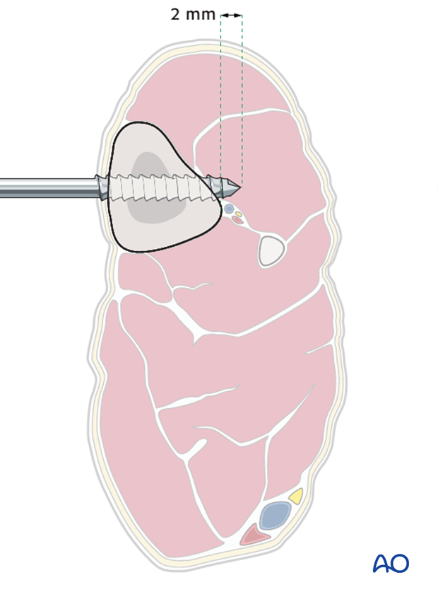
The illustration shows the appropriate pin placement centered in the bone.
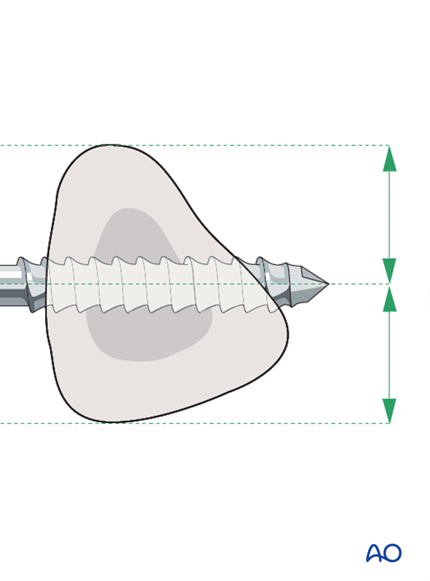
The illustration shows the appropriate pin placement centered in the bone protruding 2mm through the trans cortices.
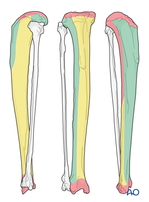
Safe corridors for the tibia
In the illustration, the green, yellow and red zone show the low, mild, and high morbidity respectively.
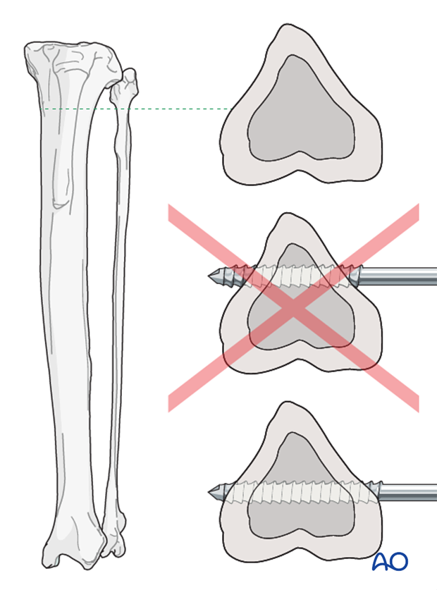
Note: In the proximal tibia, the cross section remains a triangular shape. Therefore, placing the pins caudally is preferable for better bone purchase.
4. Placement of clamps and connecting rods
The first pins inserted are the ones furthest from the fracture.
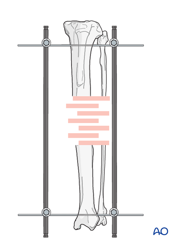
The frame is used to align the fracture. Alternatively, a distractor device can be used for this purpose.
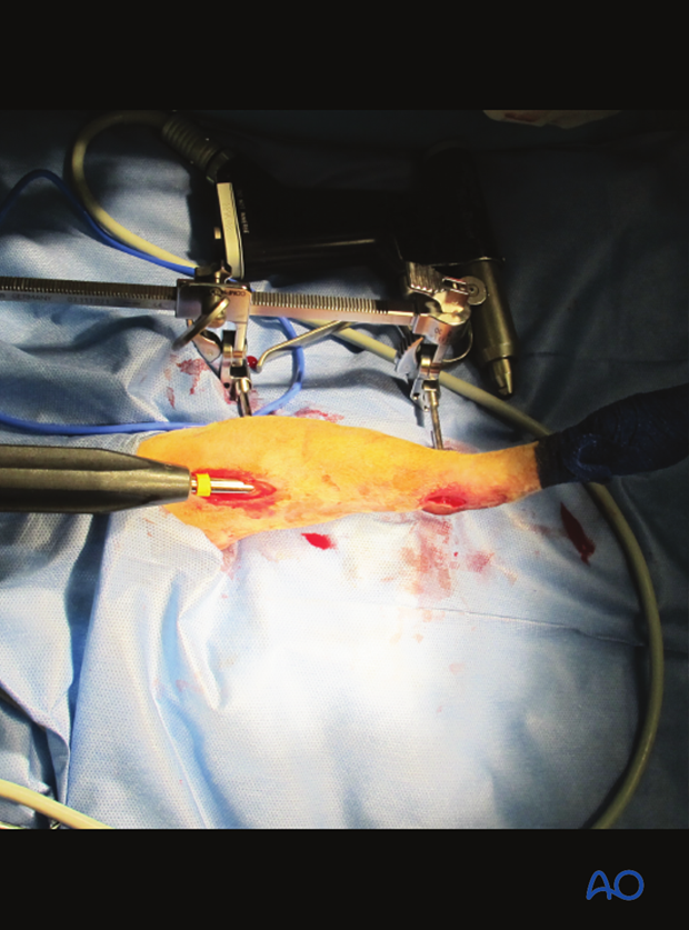
The remaining pins are inserted.
Note: One pin in each segment needs to be placed near the fracture line.
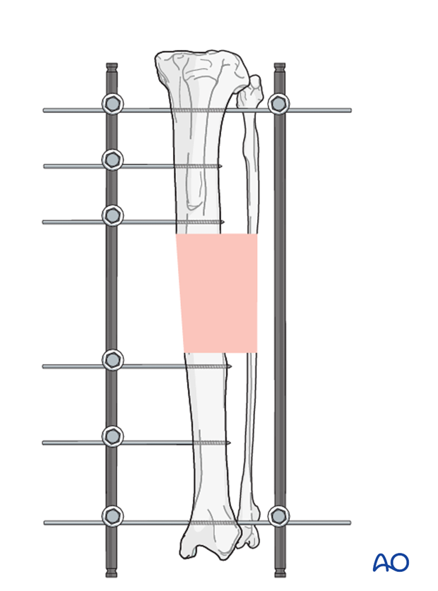
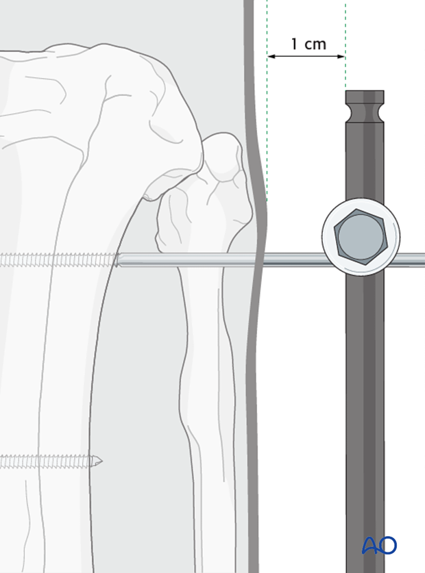
Note: The clamps and connecting bars should be approximately 1cm away from the skin. The soft tissues will swell and be in contact with the fixator if placed any closer.
5. Aftercare
Phase 1: 1-3 day after surgery
The aim is to reduce the edema, inflammation and pain. Integrative medical therapies, anti-inflammatory (note in the cat that many are toxic; only use drugs labeled for cats) and analgesics medications are indicated.
Sterile sponges should be placed in between the skin and connecting bars to protect the pin tract wounds.
The pin-skin interface should be cleaned daily.
In most cases, 10-20 minutes of ice therapy is recommended every 8 hours, but maybe challenging in cats.
Phase 2: 4-10 days after surgery
The aim is to resolve the hematoma, the edema and control the pain, as well as to prevent muscle contracture. Analgesic medications may still be needed. Anti-inflammatory medications in the cat are not labeled for continued use after a few days and should be avoided. Rehabilitation and integrative medical therapies can be used. Once the pin tract wounds are closed, the bandage can be downsized to only cover the ESF clamps and bars.
If the cat is not starting to use the limb within a few days after surgery, a careful evaluation is recommended.
10-14 days after surgery the sutures are removed.
Phase 3: 10 day-bone healing
Radiographic assessment is performed every 4-8 weeks until bone healing is confirmed.
Patients with external fixators should be checked every 7-10 days for pin track infection and fixator stability.
Disassembly or staged disassembly
When there is evidence of good callus formation, staged disassembly of the construct can be considered.
Complete removal of ESF is indicated once the fracture is healed.


