

Complications with distal radial and ulnar fractures
1. General considerations
The most common complications following repair of distal radial fractures are:
- Nonunion
- Osteomyelitis
- Malalignment and malunion
- Growth deformities due to trauma to the distal ulnar physis
- Plate failure
- Antebrachiocarpal joint degeneration in case of articular fracture
- Stress shielding
2. Nonunion
Nonunion in the distal radius is common in toy breeds. It results from a combination of biological and mechanical factors that interfere with the bone-healing process. In toy breeds, this is more pronounced since these dogs have a decreased vascularization of the distal radius and tiny or absent medullary canals.
Improper handling of the muscles and soft tissue, combined with poor surgical technique and suboptimal implant selection, can lead to delayed union or nonunion in the radius.
Linear external fixators are reported to have a higher incidence of nonunions following treatment of distal radial and ulnar fractures in toy breeds.
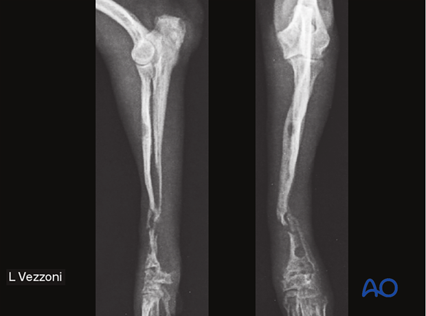
The x-rays show a radioulnar atrophic nonunion in a 4-month-old Pomeranian dog after internal fixation.
3. Osteomyelitis
In the presence of an open fracture or iatrogenic contamination, osteomyelitis can occur in the radius and ulna. The risk is particularly high in this region because of poor soft-tissue coverage.
In toy breeds the decreased vascular density may also increase the risk of infection.
4. Malalignment with malunion
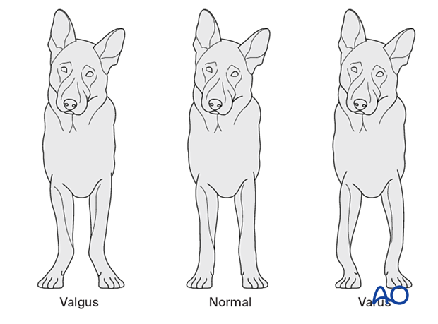
Rotational malalignment can occur in comminuted fractures due to a lack of recognizable landmarks that aid the surgeon in proper limb alignment. Valgus deformity in distal fractures may be caused by excessive retraction of the extensor tendons during fracture stabilization.
Mild loss of length or a moderate malalignment on the sagittal plane (procurvatum or recurvatum) does not affect the patient’s functional outcome. In contrast, rotational malignment and malalignment in the frontal plane (varus or valgus) can severely compromise limb function.
Limb alignment can be assessed by clinical evaluation using the flexion-extension axis of the carpus and elbow as a reference, and intraoperative fluoroscopy or x-rays may also be used.
5. Growth deformities due to trauma to the distal ulnar physis
Trauma to the distal radius is often combined with trauma to the distal ulna, which cannot be assessed at the time of injury but should be verified with a radiological recheck 2–3 weeks after.
For Salter-Harris type V fractures, premature closure of the physis of the distal ulna can occur. The radius still grows (has growth potential), whereas the ulna stops growing, causing valgus deformation, outward rotation, and procurvatum.
More rarely, the same type of injury may affect the radial growth plate, resulting in premature closure of the physis, short radius, and elbow incongruity.
6. Plate failure
When a plate is too weak, cycling load may lead to implant breakage or excessive acute load may lead to plastic deformation of the plate.
7. Antebrachiocarpal joint degeneration in case of articular fracture
In articular fractures, variable degrees of osteoarthrosis may develop in the antebrachiocarpal joint, even when adequate reduction and fixation are performed.
Lack of anatomical reduction in the articular surface, always leads to severe joint degeneration.
8. Stress shielding
Implant-induced osteoporosis may develop due to reduced mechanical stress on the bone when too stiff implants are used. This condition is typical of toy breed dogs because of the anatomical structure and blood supply.
To avoid this, careful implant selection and serial radiographic monitoring are required. In case of early detection of implant induced osteoporosis, this should be treated by staged dynamization of the implant, removing the screws closer to the fracture line thus increasing the working length of the plate and decreasing the stiffness of fixation.
In some cases, complete removal of the implant may be required as a final stage.


