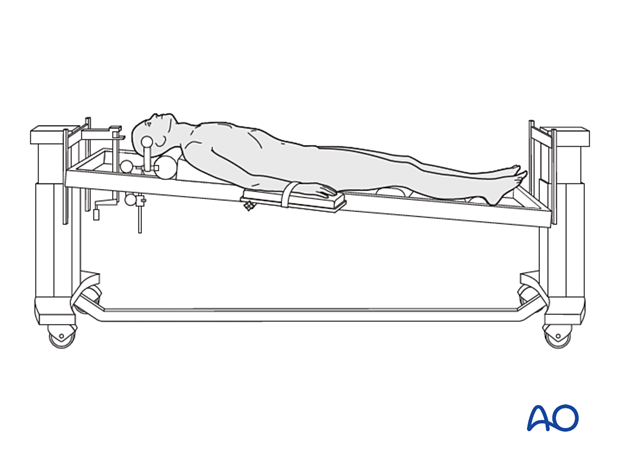
Supine position for approaches to C3–C7
1. Positioning
The patient is placed on a radiolucent table or a Maquet table in a supine position.
The neck is put into a slight hyperextension by placing a pillow under the shoulders.
An additional small pillow is placed under the neck to prevent the cervical spine from moving when the anterior spine is manipulated.
Cervical traction using Gardner-Wells tongs may be utilized.
When lower cervical levels need to be seen in lateral imaging, adhesive straps are used to pull the shoulders downwards.
Place the patient in a slight reverse Trendelenburg position to decrease the venous pressure at the surgical site. A pillow is placed under the knees to prevent the patient from sliding down the table.
A C-arm can be placed to allow for intraoperative fluoroscopy.

2. Anesthesia
With highly unstable cervical tumors, either an awake or fiberoptic intubation should be performed. In patients with severe spinal cord compression, it is essential to avoid hypotensive anesthesia, and the mean arterial blood pressure should be maintained above 80 mmHg.
3. Preoperative antibiotics
Antibiotics should be administered 30-60 minutes prior to the incision.
A cephalosporin antibiotic with good Gram-positive coverage is generally recommended.
4. Spinal cord monitoring
Spinal cord monitoring is optional and should be considered in cases with severe spinal cord compression or when spinal cord manipulation is anticipated.
5. Fluoroscopy/x-ray control
The incision can be planned based on the lateral x-ray.
An intraoperative CT scan can be used together with spinal navigation. This can assist in instrumentation placement, osteotomy cuts, and tumor delimitation.


