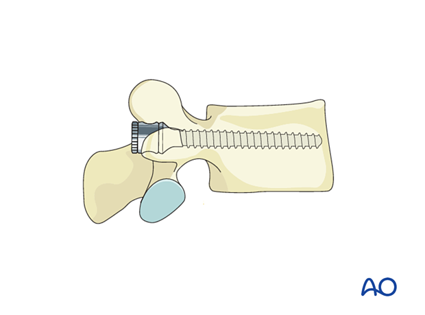
Pedicle screw insertion in the thoracolumbar spine
1. Preparation
Once the spine is exposed, the appropriate levels of fixation are confirmed with the image intensifier.
2. Pitfall: Unstable injuries
In unstable injuries, the segments above and below the level of injury may have a different orientation of pedicle trajectory due to displacement and rotation.
In severely unstable injuries, care must be taken not to cause more displacement while using the pedicle awl.
Whenever necessary, image intensifier confirmation of the starting point and trajectory must be obtained.
Preoperative assessment of pedicle integrity must be done for all levels of instrumentation.
3. Entry Points
Lumbar spine
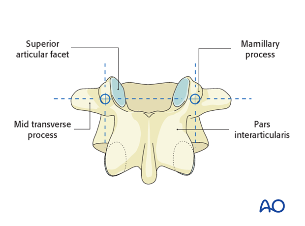
The entry point of the pedicle screw is defined as the confluence of any of the four lines:
- Pars interarticularis
- Mamillary process
- Lateral border of the superior articular facet
- Mid transverse process
Thoracic spine
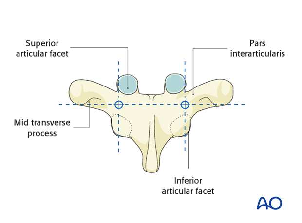
The entry point of the pedicle screw for the lower thoracic segments is defined after determining the intersection of the mid portion of the facet joint and the superior edge of the transverse process. The specific entry point will be just lateral and caudal to this intersection.
The entry point tends to be more cephalad as you move to more proximal thoracic levels.
Landmarks:
- Lateral border of the superior articular facet
- Lateral border of the inferior articular facet
- Ridge of the pars interarticularis
- Mid transverse process
4. Opening of the cortex
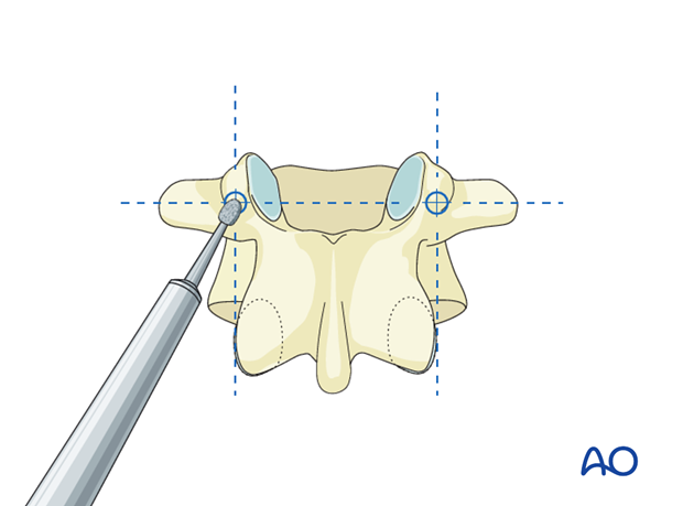
Open the superficial cortex of the entry point with a burr or a rongeur.
5. Cranial-caudal angulation
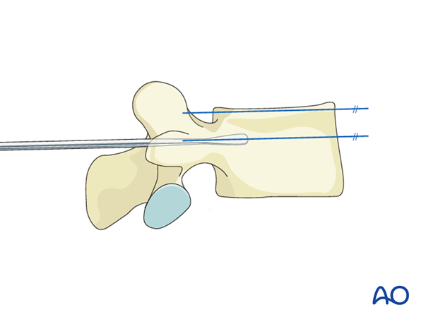
A pedicle probe is used to navigate down the isthmus of the pedicle into the vertebral body. The appropriate trajectory of the pedicle probe in the cranial-caudal direction is achieved by aiming for the contralateral transverse process, thereby aiming to be parallel to the superior endplate.
6. Medio-lateral inclination
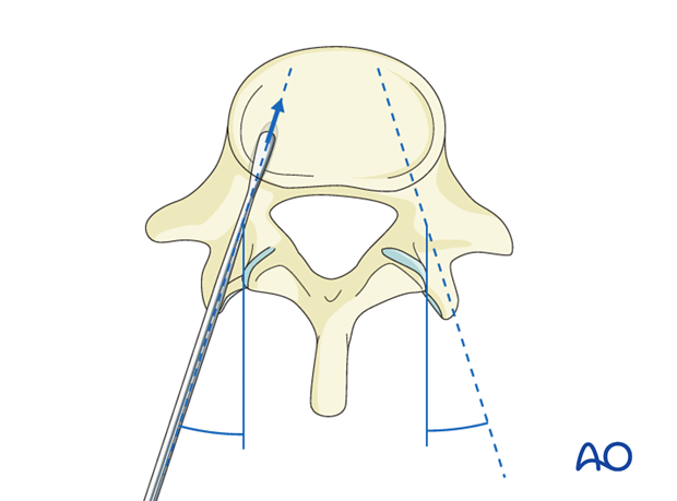
The medio-lateral inclination will depend on the rotation of the vertebra and the location, up to 45° in L5 or 0° in T5. The main goal is to avoid medial penetration of the spinal canal superficially and lateral or anterior penetration of the vertebral body cortex at the depth of insertion. Ideally, the two screws should converge but stay entirely within the cortex of the pedicles and body.
7. Screw insertion
Once the pedicle track has been created, it is important to confirm a complete intraosseous trajectory by pedicle and body palpation using a pedicle-sounding device. At any point in the process, radiographic confirmation can be obtained.
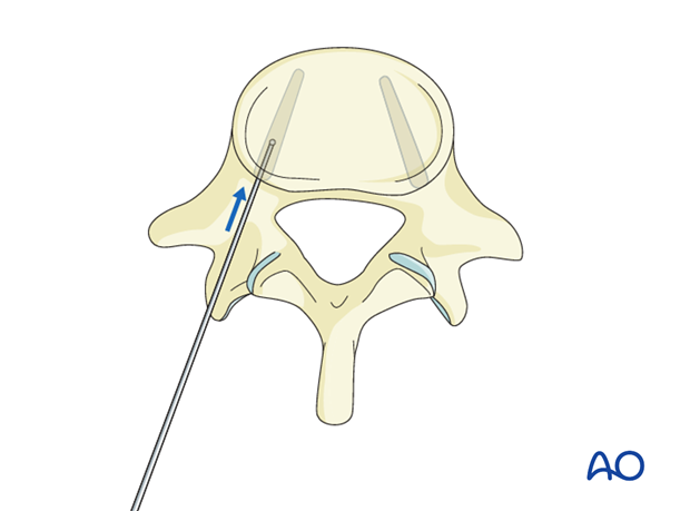
A screw of appropriate diameter and length is carefully inserted into the same created trajectory.
