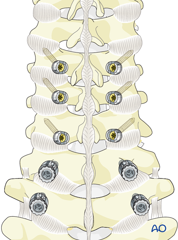
Lateral mass screw insertion (Magerl technique)
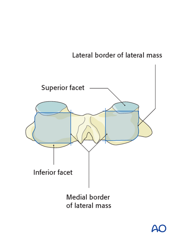
1. Anatomical landmarks
The following anatomical landmarks are identified:
- Lateral border of the lateral mass
- Medial border of the lateral mass
- Superior facet
- Inferior facet
These landmarks form a square.
2. Screw entry point
The square outlined by the landmarks is divided by a horizontal and a vertical line.
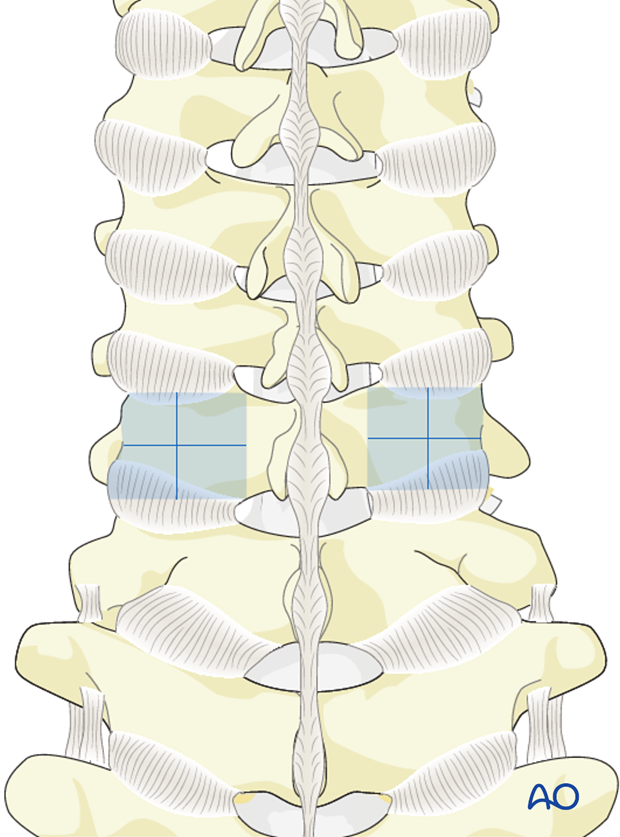
The screw entry point is located 2 mm medially and cranially to the center of the articular mass.
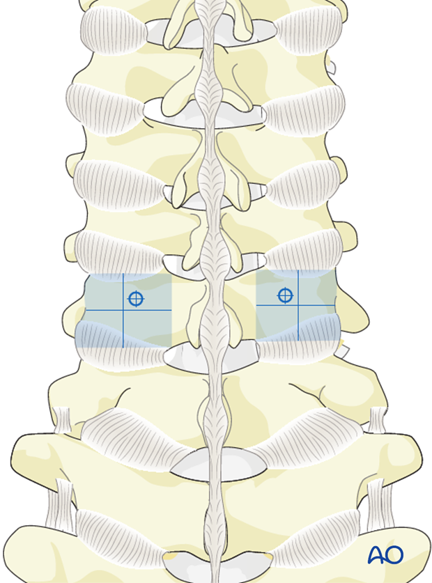
3. Opening of the cortex
Penetrate the cortex with a thin burr or an awl.
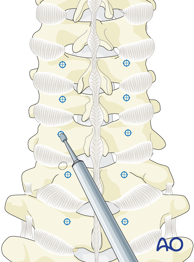
4. Mediolateral angulation
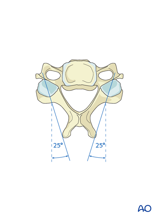
The drill trajectory should be aimed 25° laterally to avoid the vertebral artery, which is located directly anterior to the entry point.
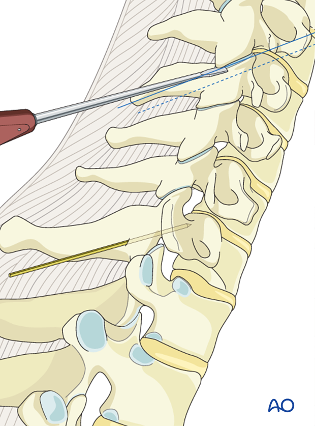
5. Craniocaudal angulation
To identify the craniocaudal angulation, a Penfield elevator is inserted in the facet joint which will be included in the fusion. The drill trajectory is then parallel to this elevator, avoiding compromise of the facet joints. Alternatively, the trajectory of the facet joints may be determined by lateral fluoroscopy.
6. Monocortical or bicortical screws?
Both monocortical and bicortical screws may be used.
Utilizing 14 mm screws will be safer, but will only allow monocortical purchase to be achieved.
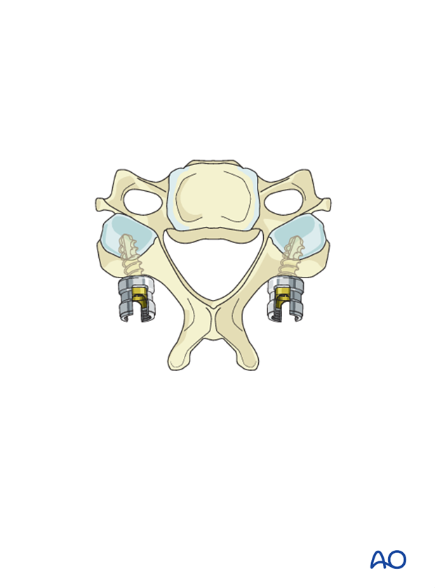
A longer screw that provides bicortical purchase will result in a more stable construct. However, the screw tip should not extend too far beyond the second cortex as it may compromise the nerve root.
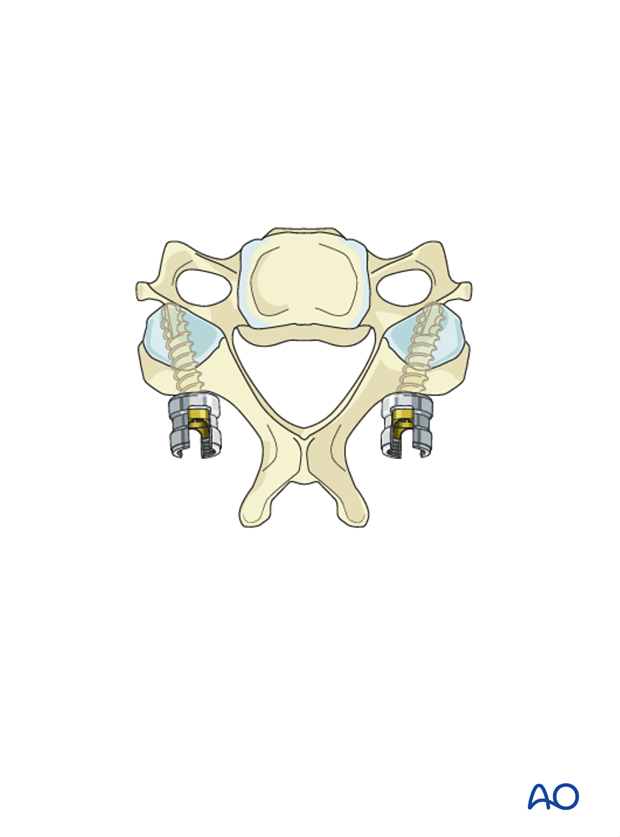
7. Drilling
If a monocortical screw is planned, the drill is set for a 14mm screw hole.
When a bicortical screw is planned, the drill is used at high speed with meticulous handling. The drill bit is advanced only for a short distance, and then pulled back before advancing again. This maneuver is repeated until the second cortex can be felt and crossed. Great care is taken to avoid over-penetration.
Alternatively, a drill stop may be used in 2mm increments. After each increase in effective drill length, the drill hole is probed to feel for penetration of the second cortex.
A screw length is selected, which corresponds to the final effective drill length required for bicortical penetration.
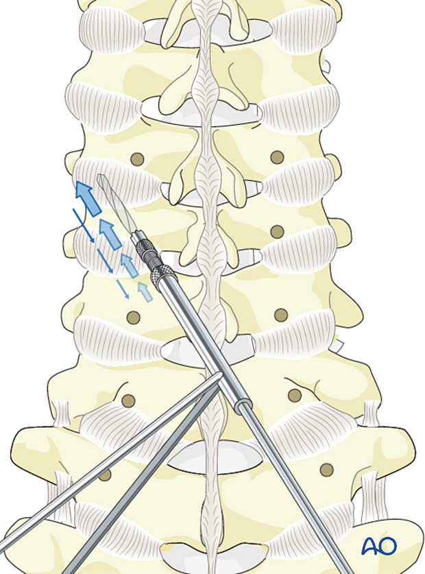
8. Screw insertion
A screw of appropriate diameter (3.5 mm) and length is carefully inserted into the trajectory that has been created.
