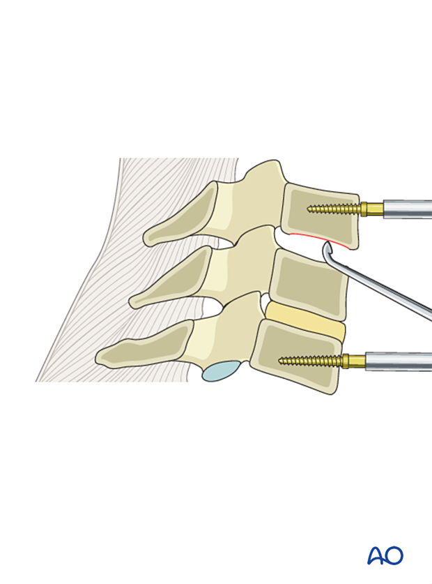
Cervical discectomy
Prior to performing the discectomy, the level is confirmed with intraoperative fluoroscopy.
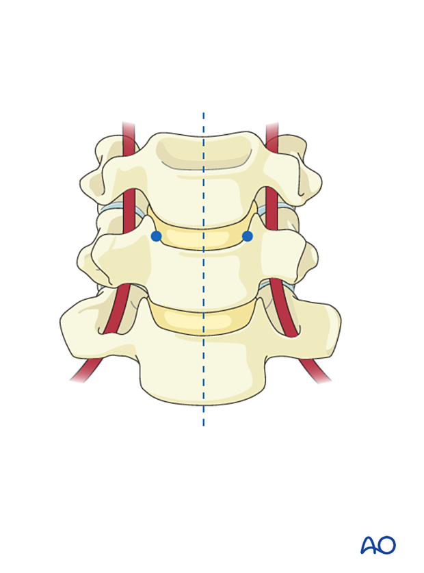
The following landmarks are often identified after the removal of osteophytes:
- Midline
- Uncovertebral process
The midline will dictate the AP orientation of the discectomy.
The uncovertebral processes dictate the lateral borders of the discectomy, establishing the safe area from the artery. This will leave a 3 mm safe zone to the normal anatomy of the vertebral artery.
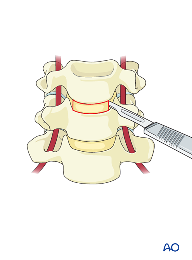
A small blade is used to open the annulus from uncinate to uncinate as close as possible to the endplates.
- To help stop a slipping blade, a suction tip is held in place in the direction the blade is cutting. Cut from each uncinate to the midline to avoid slipping laterally.
- To prevent accidental durotomy and spinal cord transection, the annulotomy should not go deeper than 11 mm.
- Cervical curettes can be used to sharp dissect the disc from the uncinates and endplates, preserving anatomy.
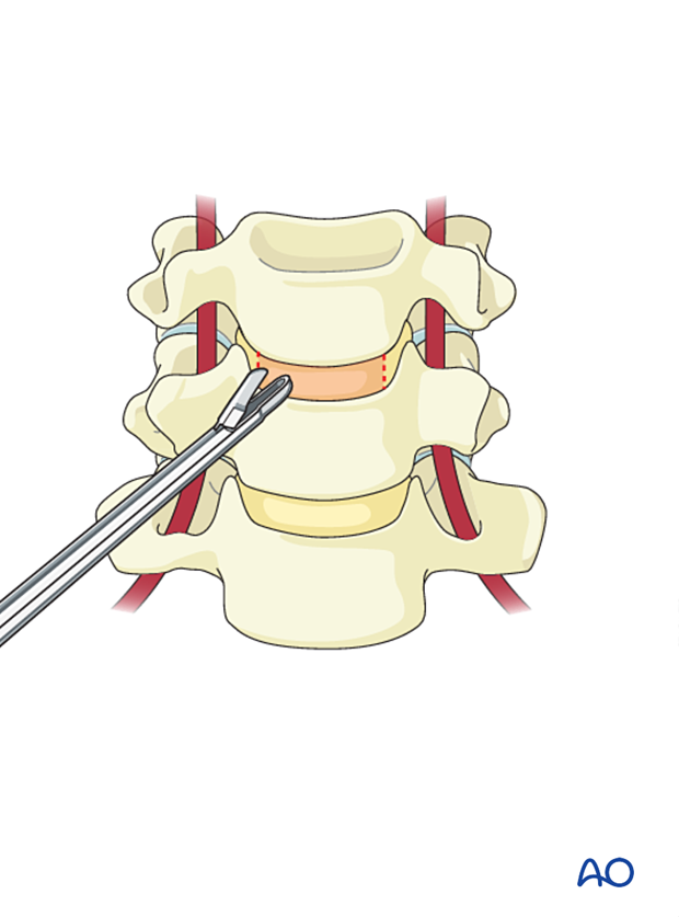
The discectomy continues using pituitary rongeur forceps to remove the annulus and small curettes to scrape the unaffected endplates.
Care is taken not to enter the tumor through the affected endplates.
The use of a distractor or an intervertebral distractor will facilitate the removal of the posterior half of the disc and visualize the posterior longitudinal ligament.
Care should be taken not to over distract.
A complete discectomy will allow good visualization of the spinal canal.
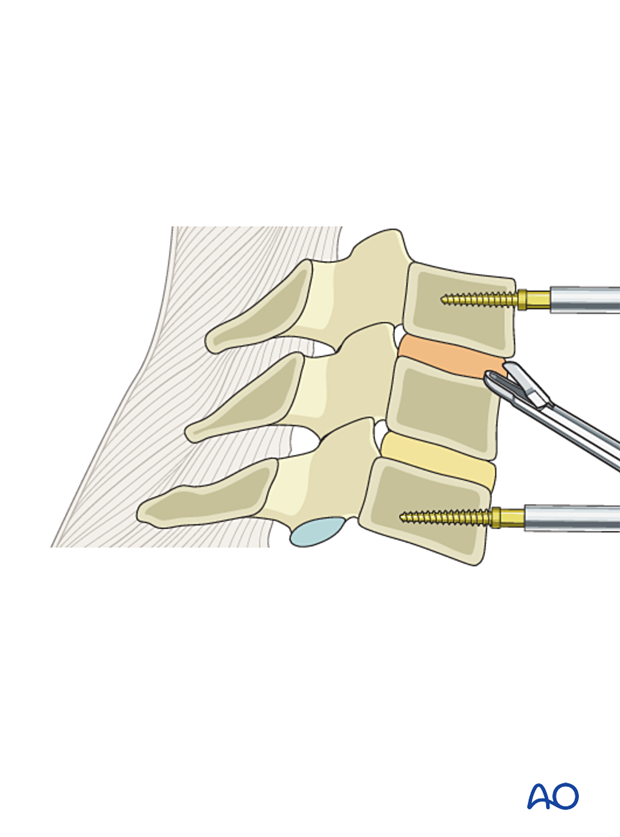
Prepare the area for the stable placement of a cage by removing cartilage from the endplates.
