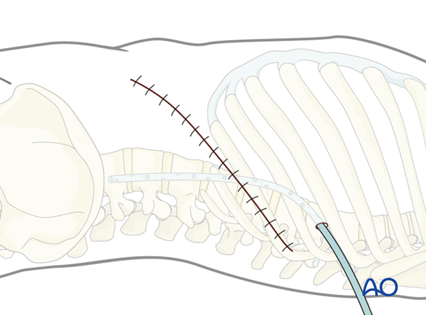
Left sided thoracolumbar junction approach (T10-L2)
1. Approach
This approach might be replaced by left sided thoracotomy or the lumbotomy, depending on the level of the vertebra
2. Skin Incision
The incision is centered over the involved vertebra. This can be verified by image intensifier.
The length of the incision depends on many factors eg, number, location and pathology, obesity of the patient, previous thoracic operations, etc.
Thanks to magnification (loupe and microscope), illumination, and table mounted retractors, the incision is typically much smaller than the traditional incision. However, to better show the anatomy, we will here show the traditional one.
It is necessary to confirm the correct level of the approach with fluoroscopy.
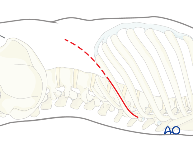
In general, the incision is made over a rib.
The rib is selected based on radiographic evaluation of the pathology and is usually the rib attached to the vertebra two levels above the involved vertebra.
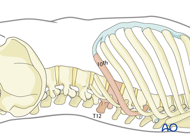
3. Exposure
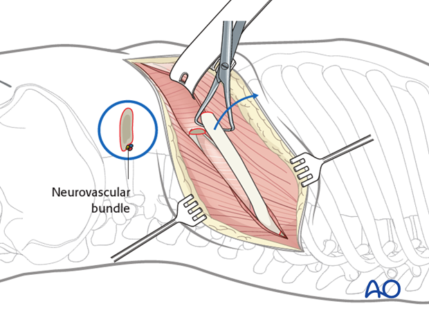
The chest muscles overlying the rib and the abdominal muscles distal to the costal cartilage are incised with cautery. The latissimus dorsi can usually be spared by retracting it posteriorly.
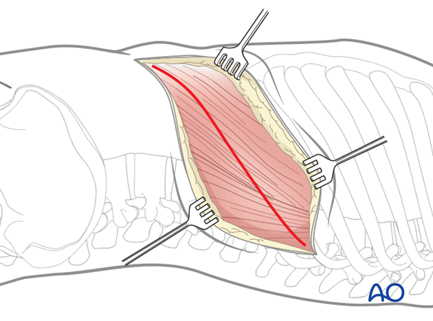
The rib is exposed subperiosteally, and if necessary, might be resected and saved for subsequent bone grafting.
Care is taken not to injure the neurovascular bundle, which lies at the caudal edge of the rib.
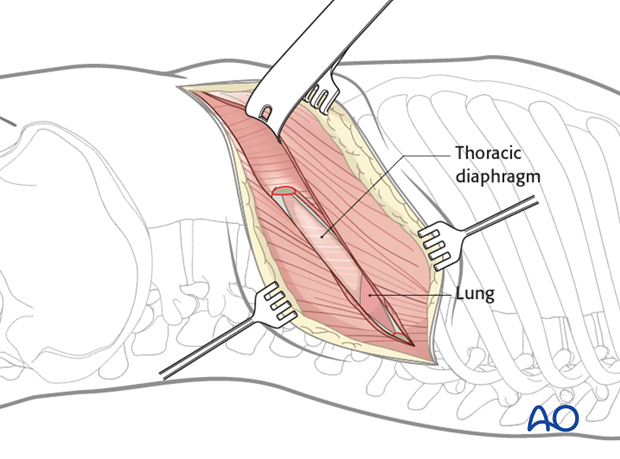
At the posterior part of the incision, the pleura parietalis is opened and the lung exposed.
Further anteriorly the diaphragm attaches to the chest wall.
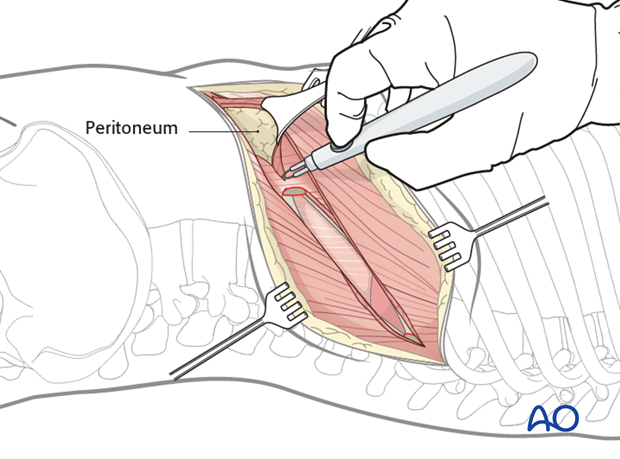
At the anterior part of the incision, the three muscle layers of the abdominal wall are split and the retroperitoneal cavity is exposed.
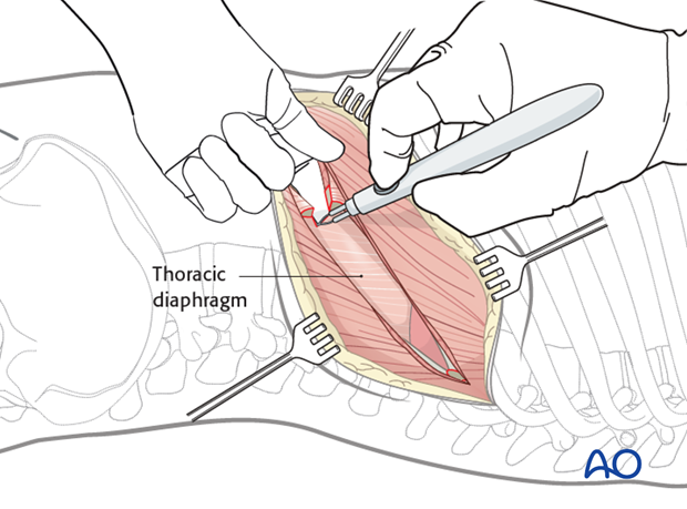
After sweeping the peritoneum off the inferior surface of the diaphragm, it is taken down from the chest wall all the way to the spine (down to the crus) leaving a 1 cm margin around the periphery for later reattachment. It may be useful to leave marking sutures every 3-4 cm to aid later reattachment.
Further steps of the approach depend on the level of the involved vertebra:
If T10-T12 have to be exposed, the principles of the left sided thoracotomy have to be followed.
If T12/L1 has to be exposed, the diaphragm has to be cut.
If L1/2 has to be exposed, the principles of the lumbotomy should be followed.
4. Access to T10-T12
To access the spine above T12, the diaphragm doesn't need to be cut and a left sided thoracotomy approach is performed.
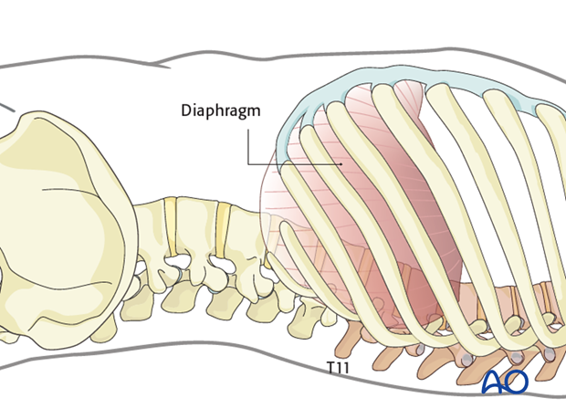
The lung is retracted anteriorly to expose the spine. It is optimal to have single lung ventilation with selective collapse of the ipsilateral lung tissue to provide exposure of the thoracic vertebral column.
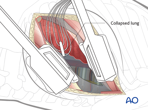
Pearl: Retraction system
It is helpful to have a table mounted retraction system to gently retract the lung and the vascular structures.
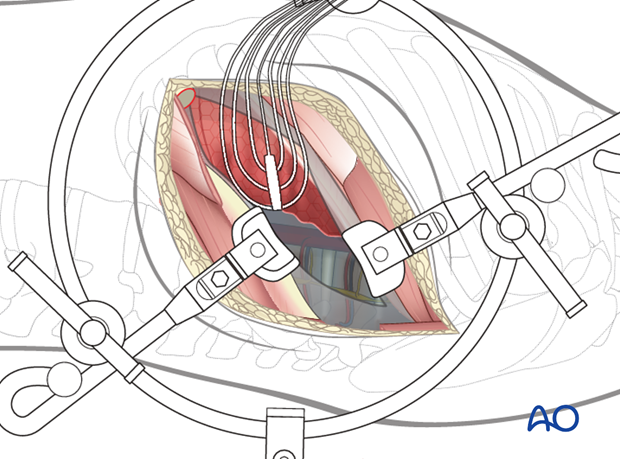
The parietal pleura should be opened longitudinally over the spine at the fracture level and a level above and below. It is easiest to begin the dissection at the discs, which will appear as avascular "hills". The "valleys" are the vertebral bodies, and the segmental vessels are in the midline.
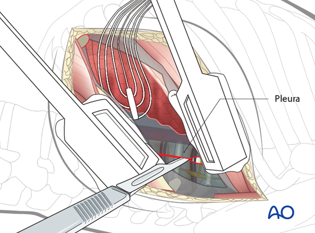
The segmental vessels at the involved level and above and below the involved level should be isolated and ligated in the mid vertebral body with sutures and/or clips.
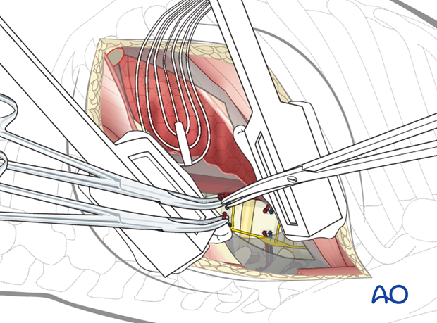
A subperiosteal exposure is then undertaken to the level of the anterior longitudinal ligaments with protection of the great vessels.
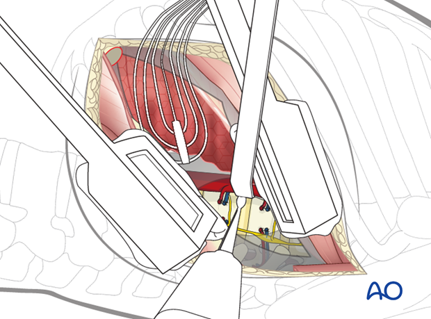
Posteriorly, the dissection should expose the heads of the ribs.
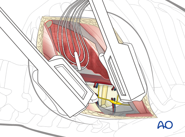
5. Access to T12/L1
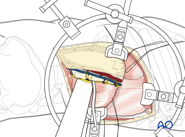
For access to the T12/L1 junction, the diaphragm has to be cut.
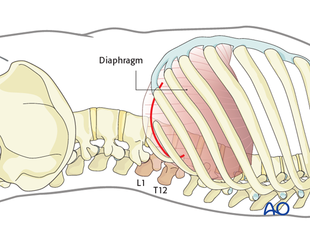
The innervation of the diaphragm comes from the phrenic nerve near the esophagus, and any muscle left standing in the periphery will be denervated. Therefore, it is important to cut the diaphragm close to the chest wall.
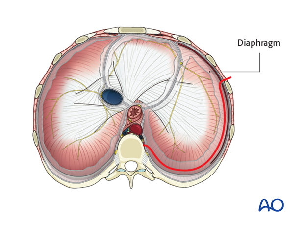
The diaphragm is then retracted anteriorly to join the retroperitoneal space and the intrapleural cavity. The lung does not need to be selectively deflated and can be easily retracted with a lung retractor and a moistened sponge for protection.
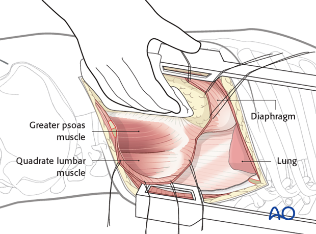
Above L1 the pleura parietalis is cut longitudinally along the lateral aspect of the spine as far proximally as one level above the involved vertebra.
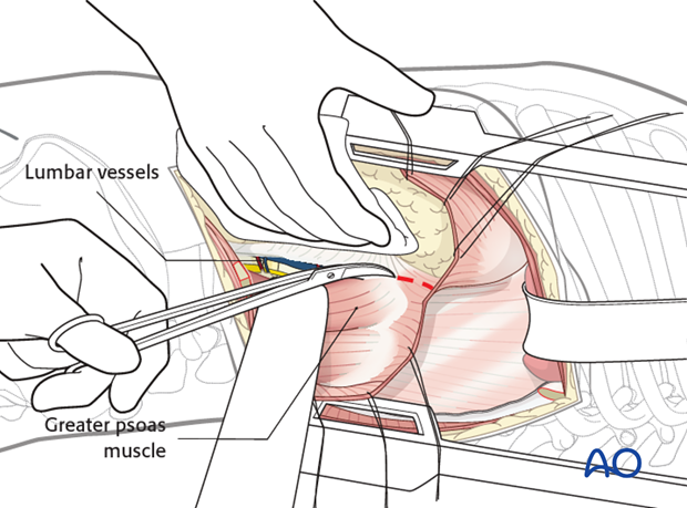
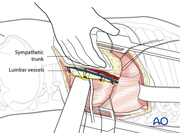
The segmental vessels that run over the vertebral bodies are exposed and ligated.
The sympathetic chain is bluntly retracted.
The spine is now exposed. Blunt retractors and swabs are placed anteriorly to protect the aorta and caval vein.
Pearl: Retraction System
It is helpful to have a table mounted retraction system to gently retract the lung and the vascular structures.
6. Access to L1/2
To expose vertebrae below L1, the peritoneum is retracted anteriorly and the lumbotomy is performed.
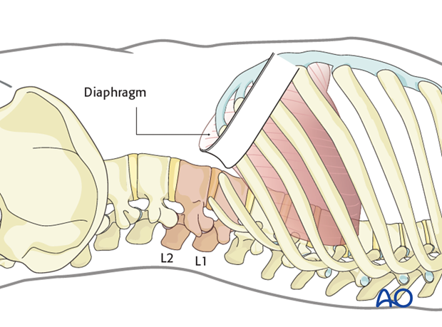
To expose L1 and L2 it is necessary to reflect the psoas muscle posteriorly.
The psoas muscle is mobilized to facilitate posterior retraction. The dissection begins on the anterior border of the psoas muscle over a disc space.
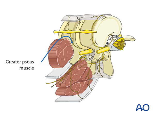
The peritoneum must be shifted away from the lateral abdominal wall until the psoas muscle is exposed. Then, the psoas muscle has to be retracted.
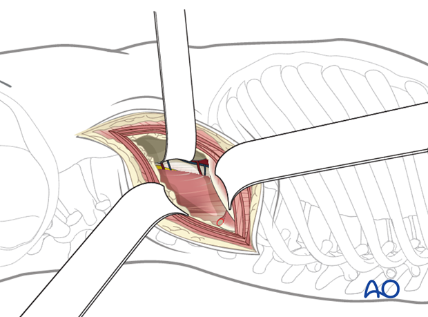
In the lumbar region, the psoas muscle is thick.
The psoas muscle is then mobilized to facilitate posterior retraction. The dissection begins on the anterior border of the psoas muscle over a disc space. The disc space above and below the involved vertebra should be exposed initially.
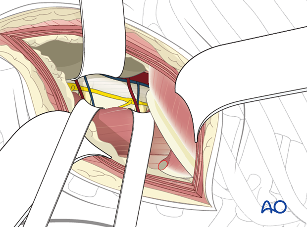
The segmental vessels above and below the involved vertebra should be isolated, ligated with sutures and clips, and divided.
Next, the segmental vessels over the involved vertebra should be isolated, ligated, and divided.
For anterior discectomies, these can be spared.
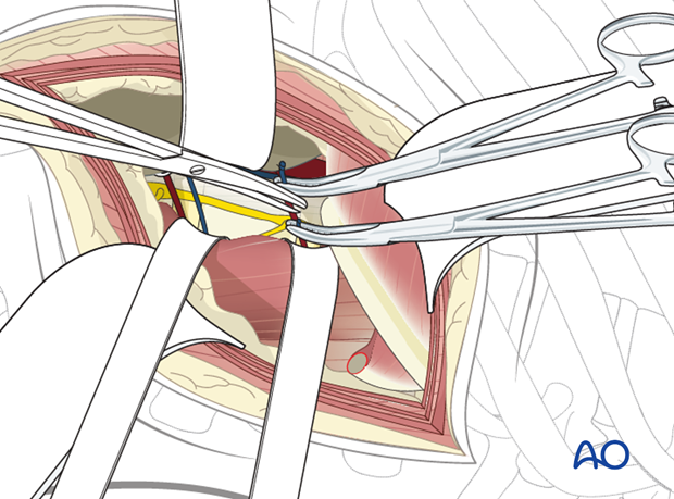
The psoas muscle can now be reflected posteriorly in a subperiosteal manner. The dissection should proceed posteriorly to the level of the pedicles. Take care not to injure the vessels in the neural foramena.
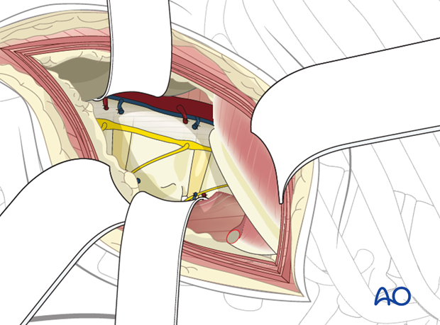
Dissection should now proceed anteriorly on the vertebral bodies above and below the involved vertebra to the level of the anterior longitudinal ligament with protection of the great vessels anteriorly. The sympathetic chain should be identified and reflected anteriorly along with the periosteum.
Next, the normal vertebral body above and below the involved vertebra should be exposed.
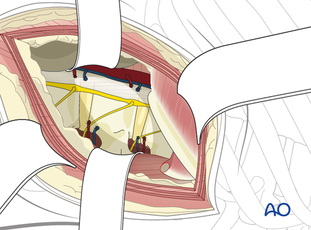
Pearl: Retraction system
It is helpful to have a table mounted retraction system to gently retract the psoas muscle, abdominal contents, and great vessels/sympathetic chain.
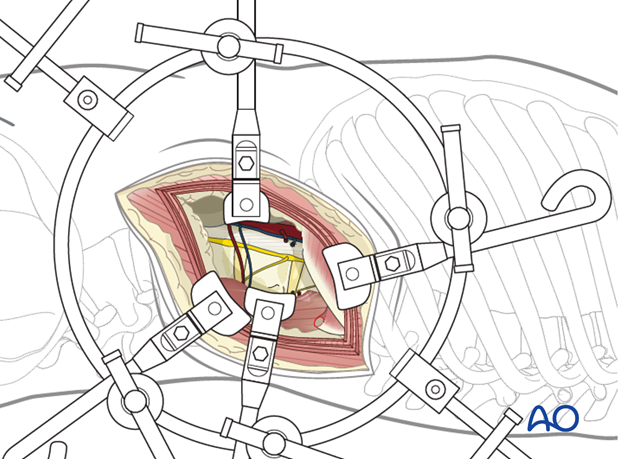
7. Closure
If possible, the parietal pleura should be sutured.
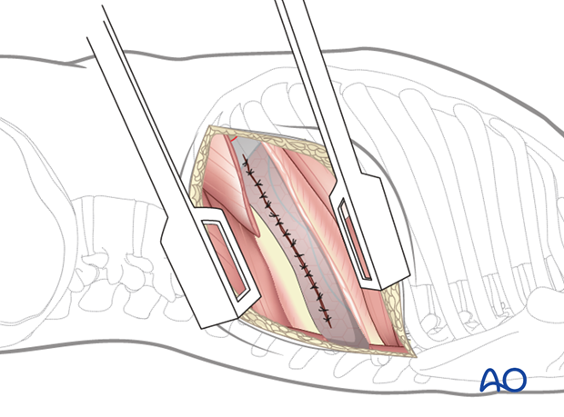
One or two chest tubes must be introduced in the mid-axillary line (some surgeons prefer an anterior drain to extract air from the chest cavity and a posteriorly placed drain for drainage of blood and fluid, others prefer just a single drain).
A retroperitoneal suction drain is optional.
The diaphragm is reattached using interrupted sutures, making sure that the pleura parietalis is approximated and preferably closed over the instrumentation.
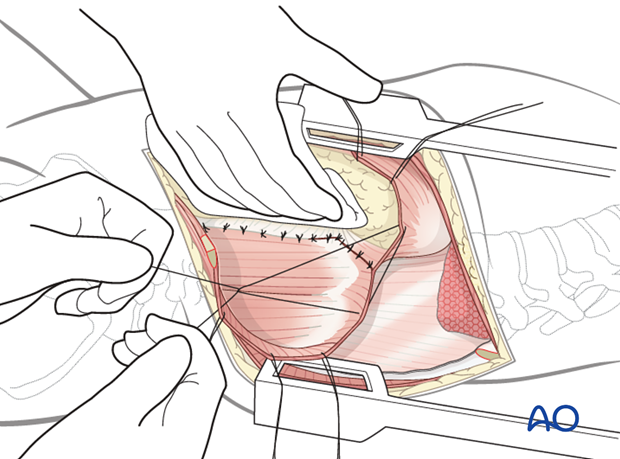
The three abdominal muscle layers are approximated.
The ribs are approximated using strong nonresorbable sutures.
Chest musculature is reattached.
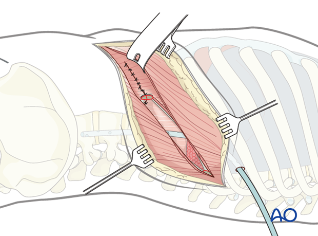
The subcutaneous tissues and skin are sutured in a layered fashion.
