Right sided thoracotomy (T3-T10)
1. Skin Incision
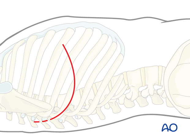
The incision is carried out from postero-superiorly around the angle of the scapula, obliquely sloping to antero-inferiorly.
The length of the incision depends on many factors eg, number, location, and classification of fractures, obesity of the patient, previous thoracic operations, etc.
It is necessary to confirm the correct level of the approach with fluoroscopy.
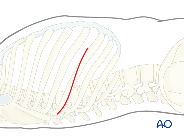
The posterior superior end of the incision should be sufficiently high to access the level above the fractured vertebra. This is best checked by image intensifier.
In general, the thoracotomy should be planned to be two levels above the fractured level.
Similarly, the most inferior part of the incision should allow access to the vertebra below the fractured vertebra.
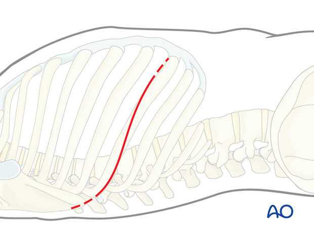
Pearl: Incision above T5
Above T5 the scapula is an obstacle. Therefore, the skin incision differs slightly and is made two centimeters below the tip of the scapula. This allows the scapula to be released from the subscapularis muscle and lifted to grant access to the appropriate intercostal space.
2. Exposure
The skin and subcutaneous tissues and overlaying muscles are incised in line, with the incision down to the rib to be exposed.
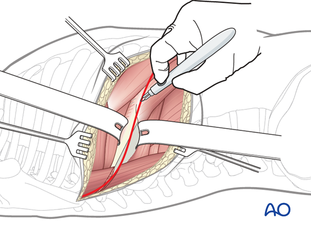
Sometimes, one rib has to be removed for access to the fractured vertebra. The rib is dissected subperiosteally and then cut from the posterior angle to as far anteriorly as possible avoiding injury to the neurovascular bundle. The rib is saved for subsequent bone grafting.
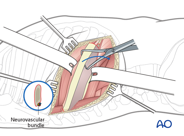
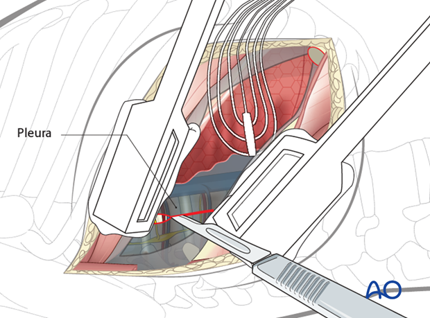
The pleura is then cut to allow entry into the chest without injuring the lung.
A rib spreader can be used to gradually open the created space.
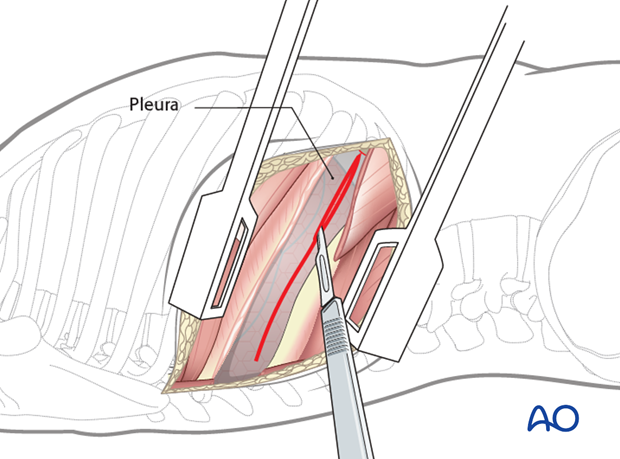
The lung is then retracted anteriorly to expose the spine. It is optimal to have single lung ventilation with selective collapse of the ipsilateral lung tissue to provide exposure to the thoracic vertebral column.
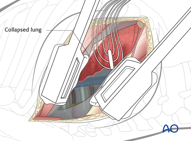
Pearl: Retraction system
It is helpful to have a table mounted retraction system to gently retract the lung and the vascular structures.
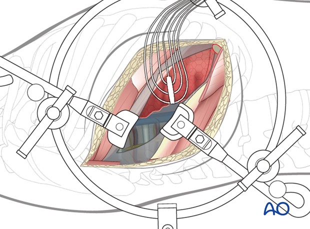
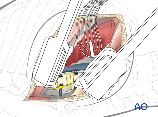
The parietal pleura should be opened longitudinally over the spine at the fracture level and a level above and below. It is easiest to begin the dissection at the discs, which will appear as avascular "hills". The "valleys" are the vertebral bodies, and the segmental vessels are in the midline.
The segmental vessels at the fracture level and above and below the fracture level should be isolated and ligated in the mid vertebral body with sutures and/or clips.
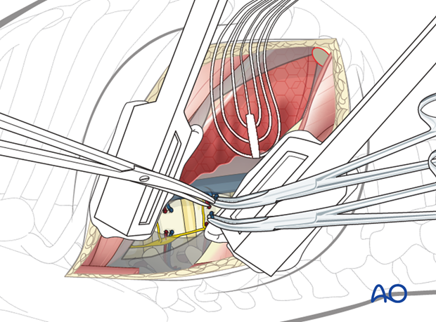
A subperiosteal exposure is then undertaken to the level of the anterior longitudinal ligaments with protection of the great vessels.
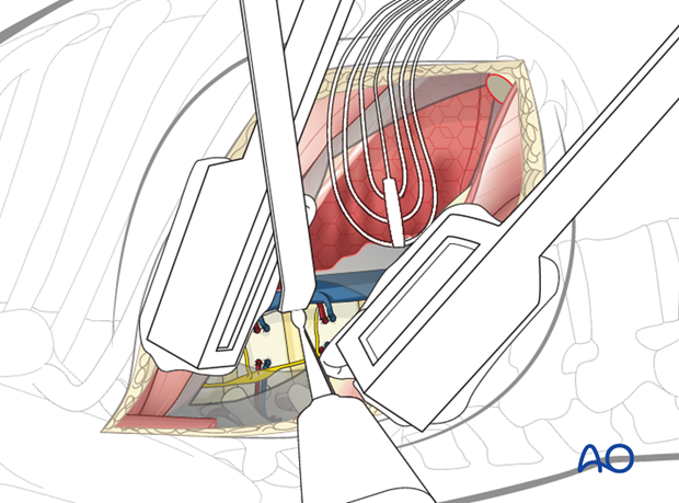
Posteriorly, the dissection should expose the heads of the ribs.
3. Closure
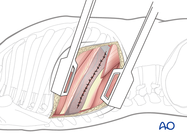
If possible, the parietal pleura should be sutured.
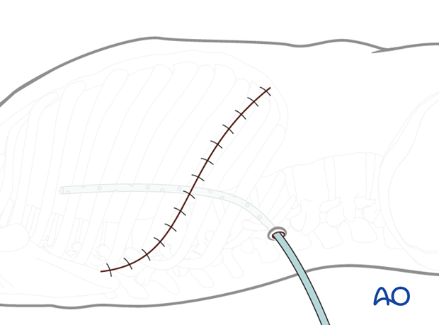
Chest drains are usually inserted via a separate stab incision.
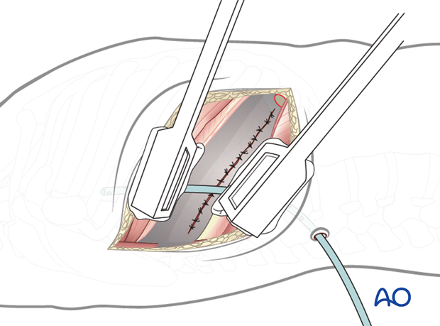
Closure is carried out in layers. Care is taken to avoid including the intercostal nerve in the suture.
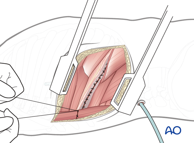
The wound is closed.
