Lumbotomy (L1-L4)
1. Skin Incision
The incision depends on the fracture level, which should be correlated with preoperative imaging.
The length of the incision depends on many factors eg, number, location and classification of fractures, obesity of the patient, previous thoracic operations, etc.
It is necessary to confirm the correct level of the approach with fluoroscopy.
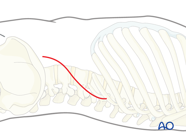
With this exposure usually three vertebrae can be easily accessed.
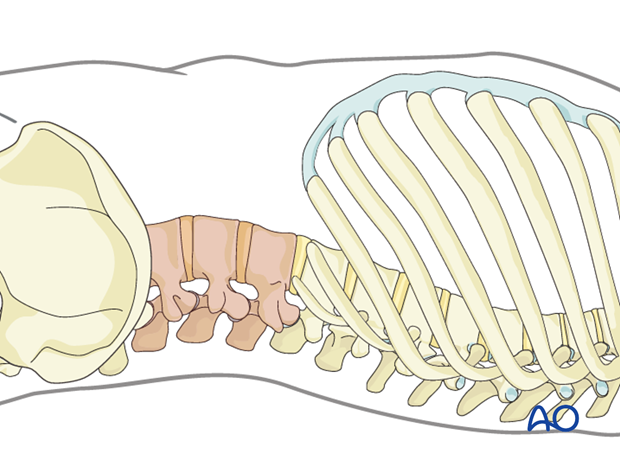
The skin is incised on the mark.
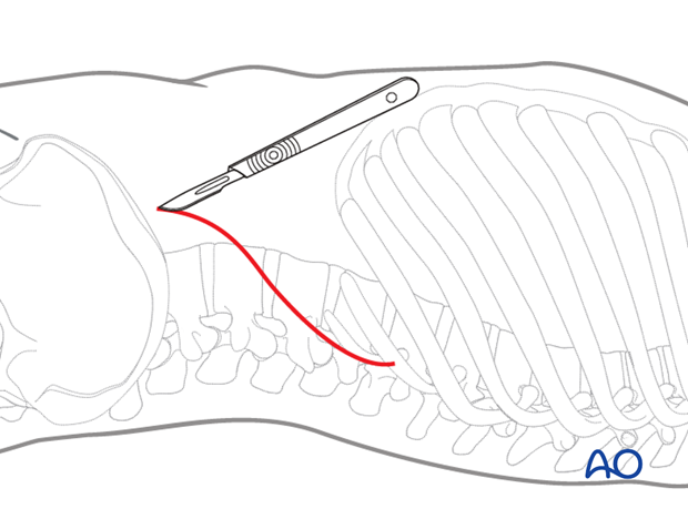
2. Exposure
There are three abdominal wall muscles. The first layer is the external abdominal oblique muscle, the second layer is the internal abdominal oblique muscle, and the third is the transverse abdominal muscle.
The subcutaneous tissue and the fascia of the obliquus externus muscle are dissected.
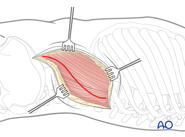
The first muscle layer is incised with cautery and retracted. The second layer is split and retracted.
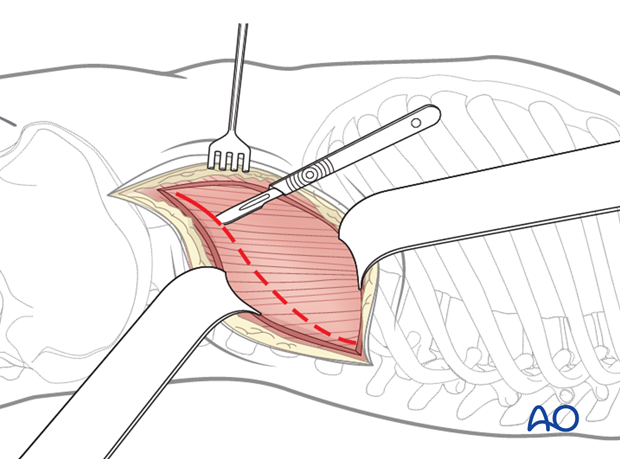
The incision should expose the distal 5 cm of the 12th rib. The rib should be exposed subperiosteally with sharp dissection and elevators.
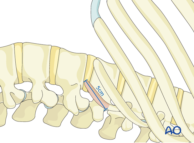
The distal 5 cm of the 12th rib should then be excised; this allows access to the retroperitoneal space.
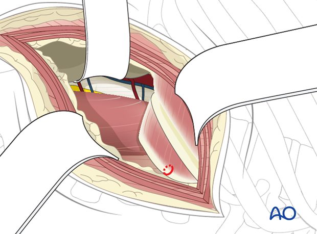
The transversalis fascia is opened with caution to avoid injury to the peritoneum that lies in the abdominal cavity.
A finger is used to split the muscle and detach it from the peritoneum to facilitate dissection.
The retroperitoneal fat is a good landmark to detect the retroperitoneal space.
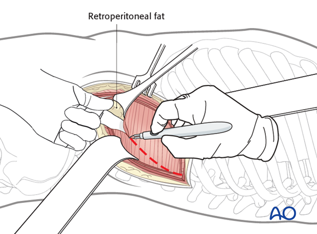
Pitfall: Injured peritoneum
If the peritoneum is injured, organs can be affected or postoperative herniation can occur.
If the peritoneum is violated, it is recommended that it be repaired directly with an absorbable suture and a blunt tapered needle.
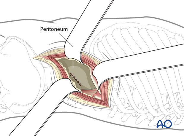
The peritoneum has to be shifted away from the lateral abdominal wall until the psoas muscle is exposed. Then, the psoas muscle has to be retracted.
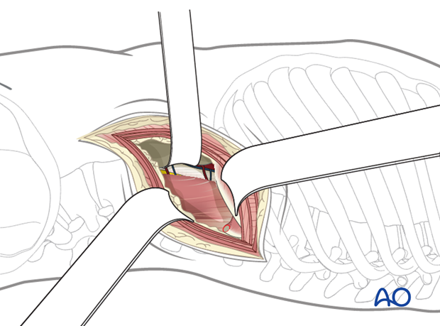
The psoas muscle is mobilized to facilitate posterior retraction. The dissection begins on the anterior border of the psoas muscle over a disc space.
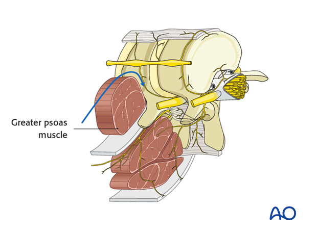
The disc space above and below the fractured vertebra should be exposed initially.
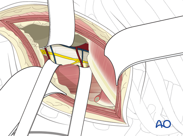
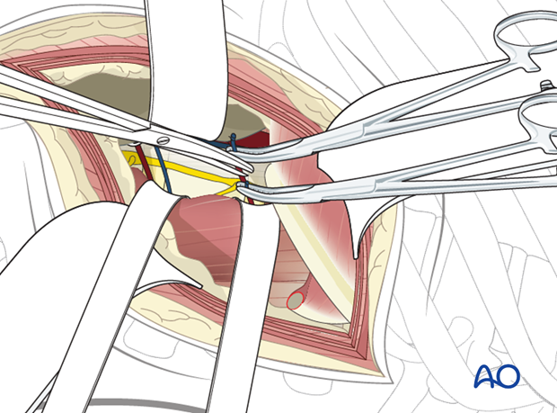
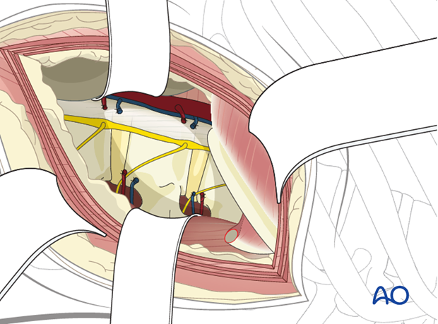
The segmental vessels above and below the fractured vertebra should be isolated, ligated with sutures and clips, and divided.
Next, the segmental vessels over the fractured vertebra should be isolated, ligated, and divided.
For anterior discectomies, these can be spared.
The psoas muscle now can be reflected posteriorly in a subperiosteal manner. The dissection should proceed posteriorly to the level of the pedicles. Take care not to injure the vessels in the neural foramena.
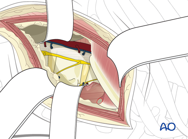
Dissection should now proceed anteriorly on the vertebral bodies above and below the fracture site and the fractured vertebra to the level of the anterior longitudinal ligament with protection of the great vessels anteriorly. The sympathetic chain should be identified and reflected anteriorly along with the periosteum.
Next, the normal vertebral body above and below the fractured vertebra should be exposed.
Pearl: Retraction system
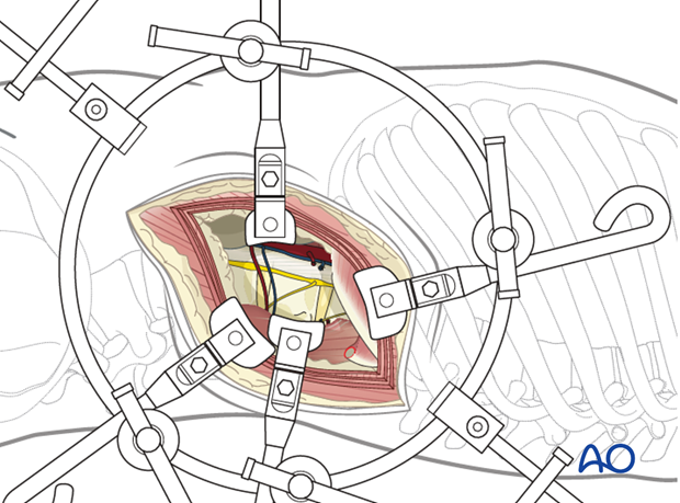
It is helpful to have a table mounted retraction system to gently retract the psoas muscle, abdominal contents, and great vessels/sympathetic chain.
3. Closure
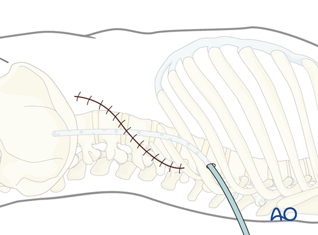
A retroperitoneal suction drain is optional.
The three abdominal muscle layers are approximated.
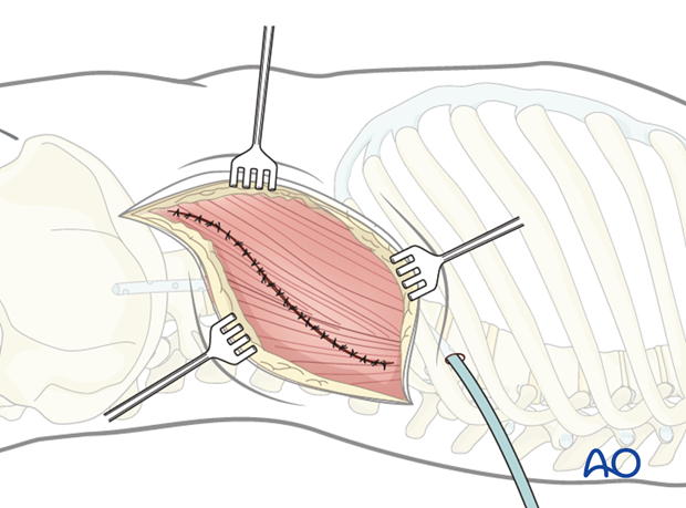
The subcutaneous tissues and skin are sutured in a layered fashion.
