Postoperative cervical deformities
1. Causes of postoperative deformities
Introduction
The most common posttraumatic cervical deformity is kyphosis. Rotational deformity and coronal plane angulation may also occur.
The most common causes are:
- Missed injuries
- Poor understanding of injury pathomechanics
- Failure of stabilization construct
- Iatrogenic destabilization
- Inadequate follow up
- Lack of resources
Missed injuries
The most common missed injuries are unrecognized ligamentous injuries (type B misdiagnosed as type A).
These generally lead to kyphosis due to loss of the posterior tension band and possibly translation caused by facet dislocation.
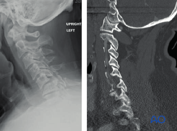
These CT-images were obtained at the time of injury. A C5-C6 ligamentous injury was unrecognized.

Three weeks post injury the ligamentous injury has manifested itself with kyphosis and displacement due to C5-C6 facet dislocation (F4).
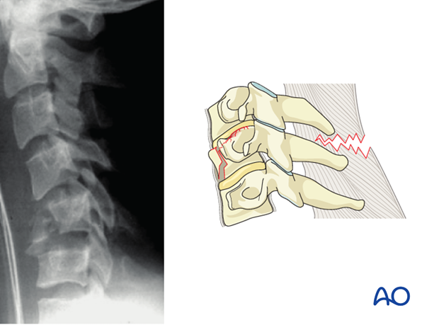
Anterior bony compression injuries (Type A2) are prone to progressive kyphotic deformity if associated with unrecognized posterior ligamentous injuries.
Unrecognized ligamentous injuries adjacent to stabilization constructs done for more obvious injuries can lead to rapid adjacent segment kyphosis.
This unilateral C4-C5 facet dislocation was treated with ACDF.
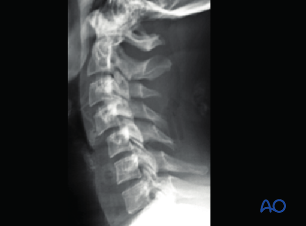
6 weeks postoperatively upright lateral radiograph shows kyphosis at the C5-C6 level.
MRI demonstrates previously unrecognized posterior ligament injury.
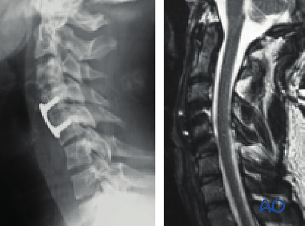
The patient required posterior stabilization involving the C5-C6 level.
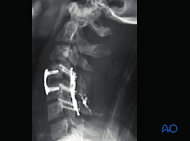
Poor understanding of injury pathomechanics
Injuries with loss of posterior tension band are likely to exhibit progressive kyphosis and need to be stabilized, especially if the anterior column is also injured.
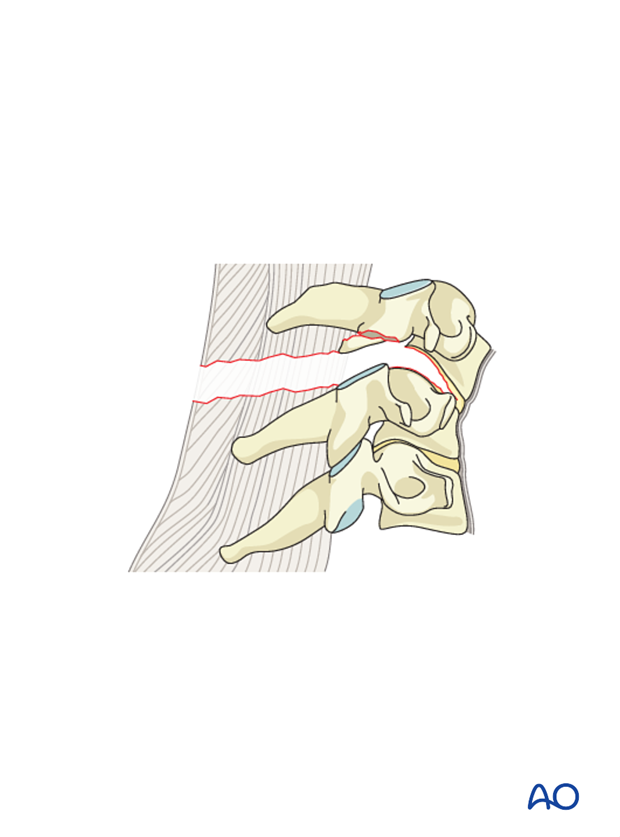
Injuries with significant anterior column compromise are at risk for failure of fixation and kyphosis after posterior stabilization alone.
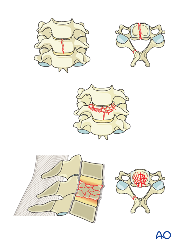
Highly unstable 3 column injuries are at risk for failure of fixation and kyphosis after anterior fixation alone.

Failure of stabilization construct
This can occur due to:
- Patient related factors such as osteoporosis or chronic disease
- Wrong choice of fixation for a specific type of injury
- Suboptimal technique
- Nonunion
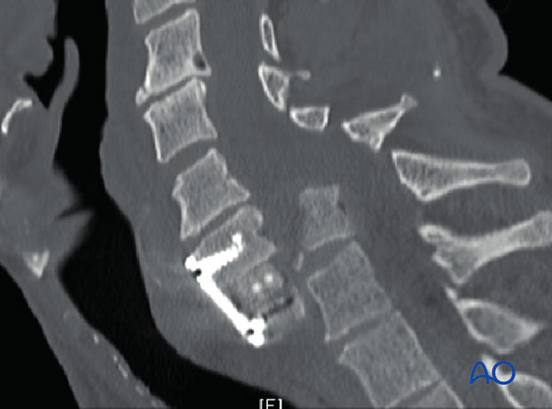
This postoperative CT shows that a C6-C7 dislocation has been left unreduced after ACDF.
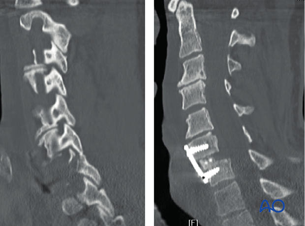
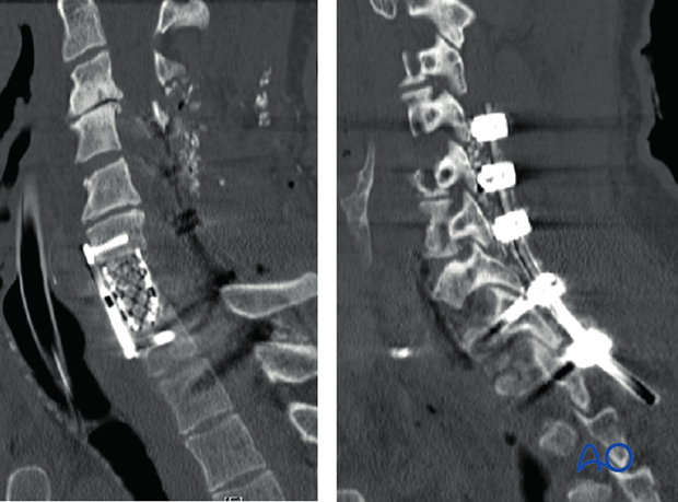
One week postoperatively the patient had new onset neck pain and quadriparesis. CT demonstrates failure of fixation.
Decompression and realignment is achieved through an anterior and posterior approach.
Iatrogenic destabilization
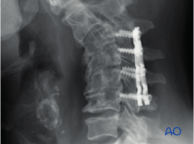
When stabilizing an injury it is essential to avoid causing injury to adjacent levels. The structures most at risk are facet capsules and interspinous ligament posteriorly.
This may lead to adjacent segment kyphposis.
Repairing the paraspinous muscles to C2 is essential to avoid cervical kyphosis.
Inadequate follow up
Any spine fracture has the potential to behave unpredictably. Unanticipated changes in alignment are most easily corrected early on.
It is therefore essential to re-evaluate both operatively and nonoperatively treated fractures radiographically and clinically at regular intervals. Initial follow up a minimum of three weeks and six weeks postoperatively is recommended.
These images show a well aligned C6-C7 unilateral facet fracture.
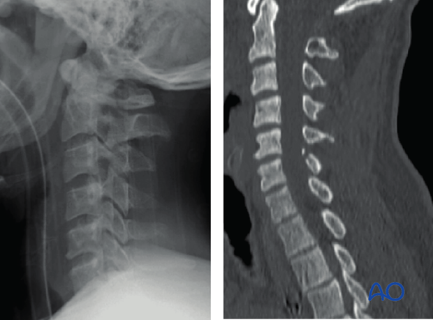
Three months post injury the patient is experiencing progressively worsening C7 radiculopathy. Radiographs show C6-C7 anterolisthesis.
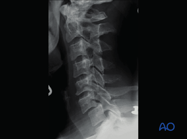
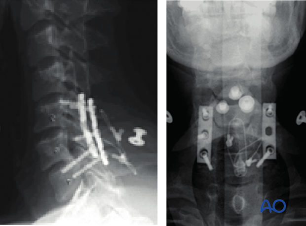
Because the deformity had become relatively stiff over time, correction required facet osteotomy, reduction with oblique wiring, and two level posterior instrumented fusion.
An alternative approach would have been two or three stage anterior-posterior approach.
2. Treatment
There are no absolute values for acceptable deformities. Whether a deformity is sufficiently problematic to warrant surgical correction is made on an individual basis and is a function of clinical judgment and the patient's specific circumstances.
The surgical approach is a function of the type, the severity, and rigidity of the deformity. We will focus on kyphosis which is the most prevalent posttraumatic deformity.
The principles involved include release of rigid deformities, correction of alignment, stabilization and reconstruction of areas of bone loss, particularly restoration of anterior column support.
Areas of nerve root compression should also be addressed as needed in the form of corpectomy, laminectomy, or foraminotomy.
The goal is to restore alignment. If correction of deformity results in an anterior column defect, anterior reconstruction with interbody spacer or cage should be included, either with anterior plate instrumentation alone or with posterior fixation.
For rigid deformities the extent of surgery depends largely on the amount of deformity correction that is required.
Posterior approach only
Posterior approach only is rarely applicable for posttraumatic kyphotic deformities. The indications include:
- Flexible deformity (this typically requires early intervention)
- Ability to achieve adequate lordosis
- No need for anterior decompression
Anterior approach only
This is rarely applicable in posttraumatic kyphotic deformities. The indications include:
- Less severe deformities
- Some degree of flexibility (no posterior osteotomy required)
- The presence of healthy bone
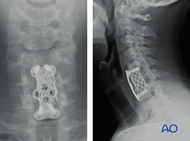
This patient with a remote history of low grade flexion-compression fracture at C5-C6 which healed in relatively mild kyphosis presented with neck pain and new onset of two level radiculopathy.
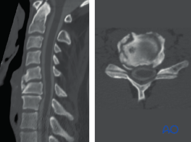
Anterior only procedure consisting of C6 corpectomy, correction of kyphosis and C5-C7 anterior fusion resulted in spinal realignment and decompression.
360 approach
Required for rigid and severe deformities. Anterior followed by posterior approach is most widely used.
The principle is to resect the apex of the deformity anteriorly through wide single level or multilevel corpectomies.
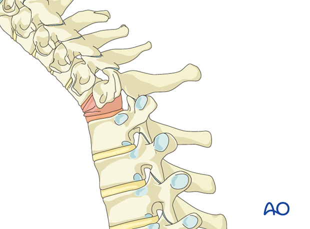
Caspar pins are used to obtain correction of the kyphosis. The amount of correction obtained is a function of the width of the corpectomy and the rigidity of the posterior spine.
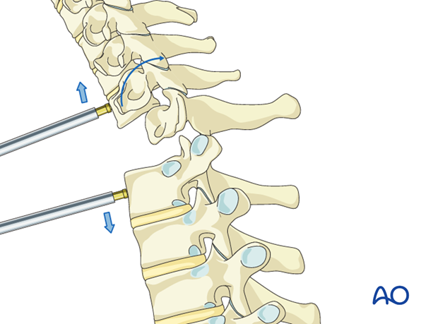
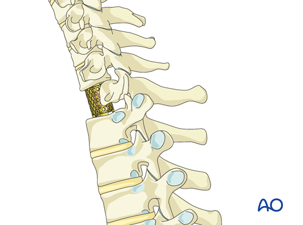
Anterior cage reconstruction may then be performed and Caspar pins are removed.
Anterior plating is rarely performed at this time as it would prevent the possibility of achieving additional lordosis during the posterior approach.
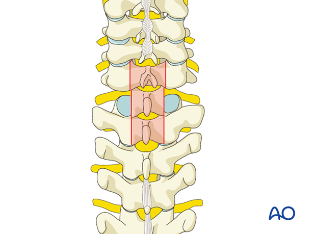
Posterior osteotomy is performed at multiple levels depending on the degree of additional correction required.
Laminectomies and foraminotomies are performed as needed for spinal cord nerve root decompression.
Interspinous wiring may be used to achieve additional lordosis if needed.
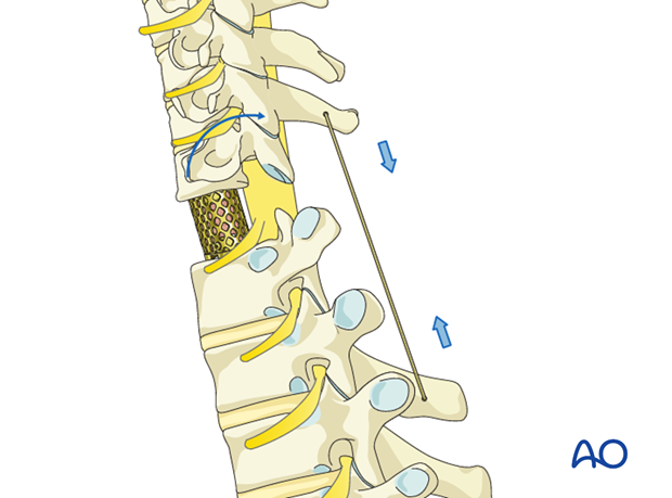
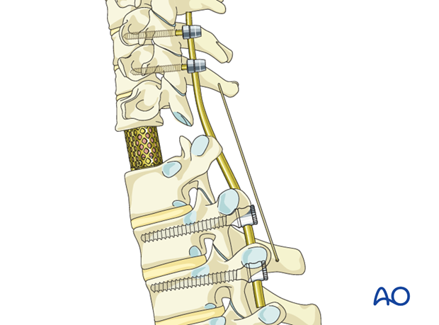
Multilevel fixation is then performed. The posterior rods are contoured according to the desired cervical alignment.
Compression is achieved across multiple levels to further accentuate lordosis.
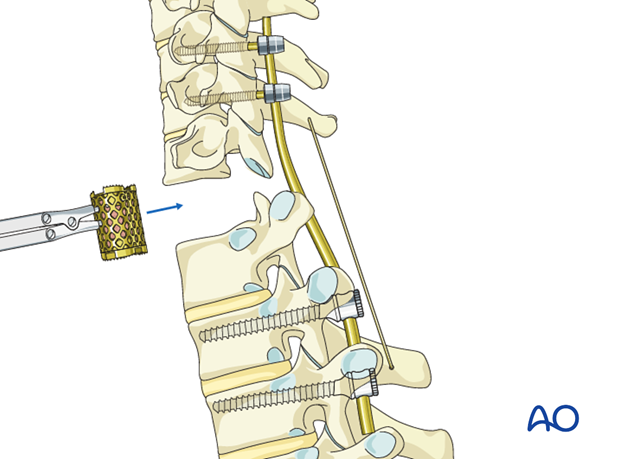
For the most severe and rigid deformities a 540 degree approach may be required. The sequence most commonly performed is anterior – posterior – anterior. The primary modification to the 360 approach is that, because sufficient deformity correction cannot be achieved from the initial anterior approach.
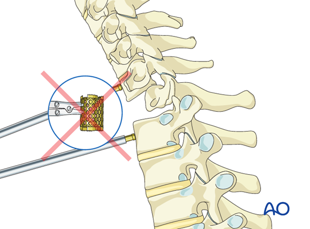
The cage reconstruction can be done only after the posterior osteotomy and instrumentation has been completed.
An alternative 540° approach follows a posterior – anterior – posterior sequence. In this circumstance the posterior osteotomies are performed first. Screws can be placed at this time, but no rods.
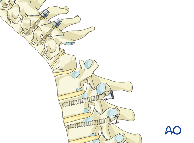
The anterior release allows for correction of the deformity because the posterior spine has already been mobilized, therefore anterior reconstruction can be performed.
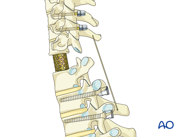
The final step is to apply rods posteriorly. Additional compression across the posterior instrumentation can be used to accentuate lordosis if necessary.
Note: Because of the multiple procedures and associated risks, the decision should be based on:
- Neurological risk or deficit progression
- Severe or progressive deformity


