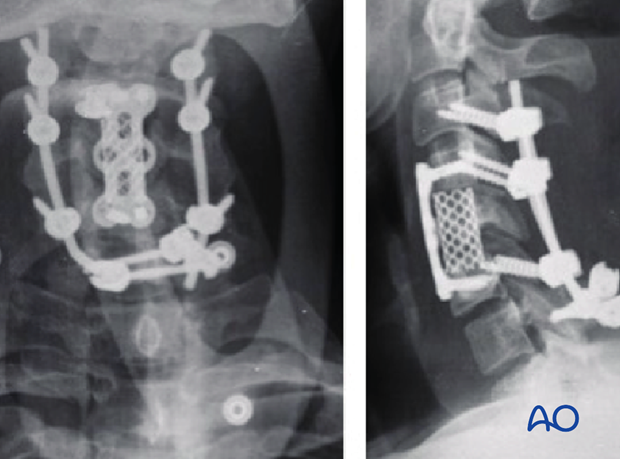
Laminar screw insertion in T1 and T2
1. General considerations
Laminar screws in T1 and/or T2 can be used as an alternative to pedicle screws, in particular when pedicles have been found to be narrow in preoperative CT scans.
Laminar screws can only be used if the posterior structures are intact. If the spinal process or the lamina are damaged, laminar screws are contraindicated.
Care must be taken if there is any damage to the pedicles since translaminar screws rely on the integrity of the pedicles transmitting forces through the vertebral body.
The appropriate screw dimensions to ensure adequate bone purchase and prevent cortical compromise should be determined from preoperative CT scans.
2. Screw entry point
Craniocaudal placement: The first screw entry point is found slightly cranial at the spinolaminar junction. It should allow for the contralateral screw to pass caudally to the first one.
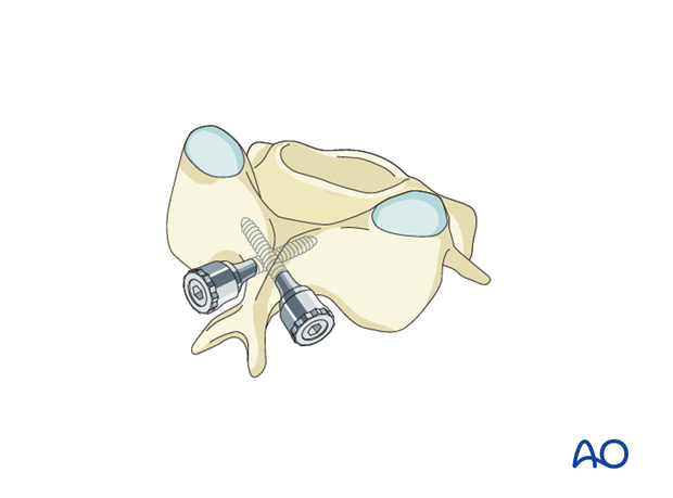
Anterior/posterior placement: The entry point should be placed in the central plane of the contralateral lamina.
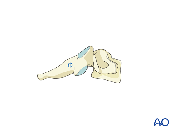
3. Preparation of screw hole
The cortex is opened using a 3 mm cutting burr.
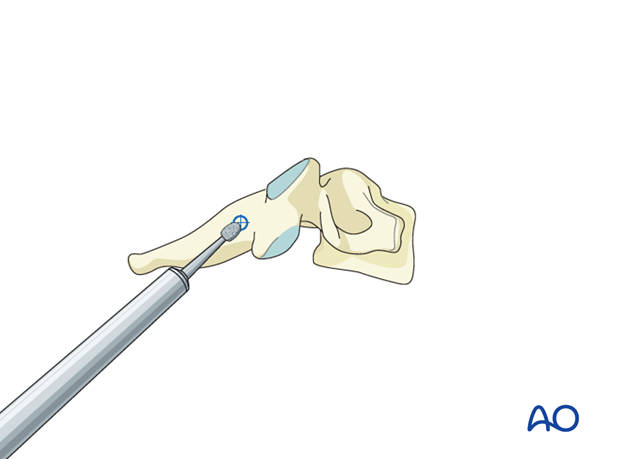
A 2 mm hand held drill is used drill the intralaminar hole.
Great care must be taken not to penetrate the non-visualized ventral laminar surface. To aid in this, the visualized dorsal surface is used as a reference to angle the trajectory slightly dorsally.
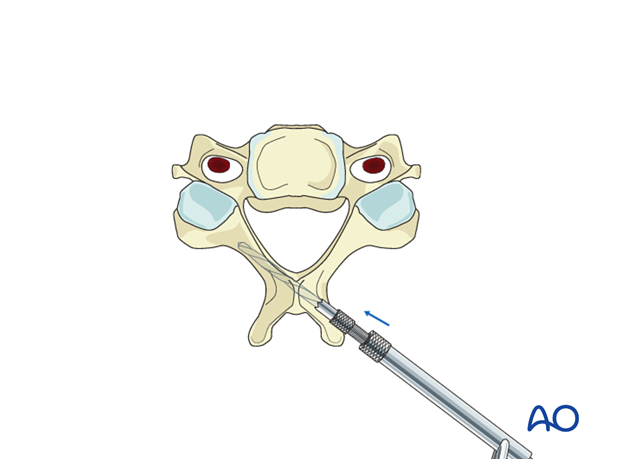
Palpate the drill hole with a ball-tipped probe to ensure the entire screw hole is surrounded by bone.
In cases where decompression has been performed above or below the T1-T2 level, watch for deflection of the dura during the probing and also later during the screw insertions.
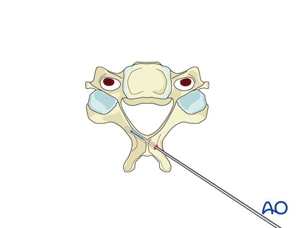
4. Screw insertion
A translaminar screw is then inserted.
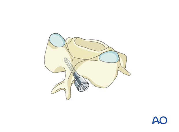
The procedure is then repeated on the contralateral side with the screw entry-point slightly caudally.
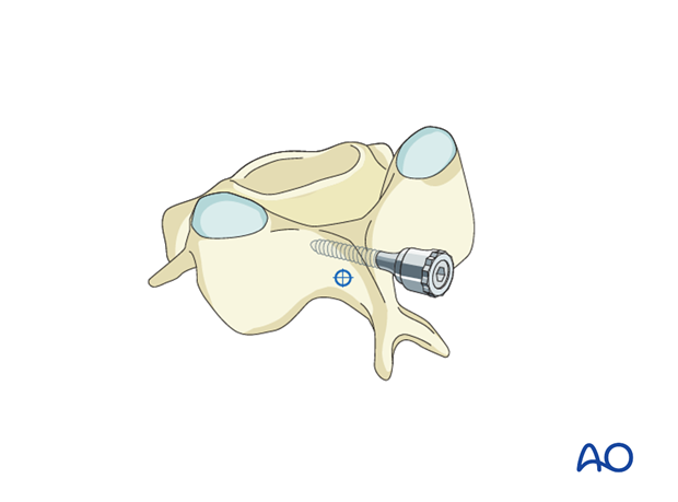
Postoperative X-ray showing C7 laminar screws.
