Anterior approach to the cervico thoracic junction
1. Introduction
This approach is used for access to the anterior C7-T1 through T1–T2 levels for decompression or reconstruction of the injured anterior spinal column. Depending on the patient's anatomy, access to lower levels may also be possible.
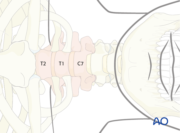
Access to the lower levels may require partial resection of the manubrium and clavicle (manubrial window).
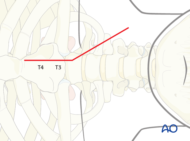
Although a formal sternotomy can be used for improved access to the upper thoracic spine below T2, this exposure can be achieved with greater ease and less morbidity using alternative posterolateral approaches.
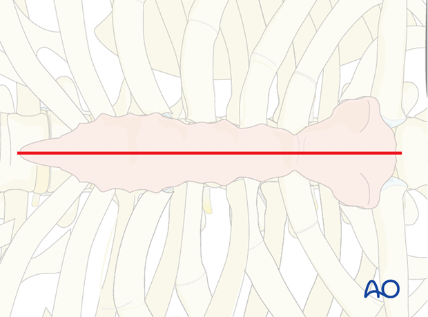
Injuries of the esophagus can be associated with fractures or occur as a complication of the approach.
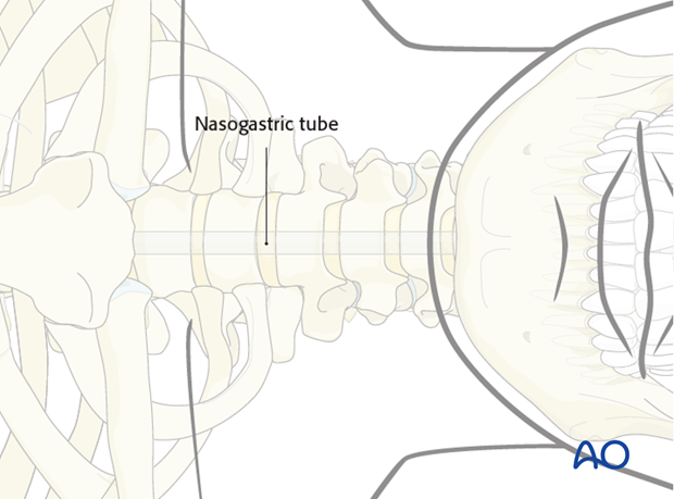
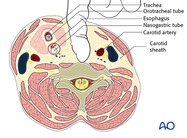
In addition to the tracheo tube a nasogastric tube should be inserted to better identify and thus help prevent accidental injury to the esophagus.
These are serious and potentially lethal complications. Consultation with thoracic or ENT surgeons should be obtained.
2. Incision
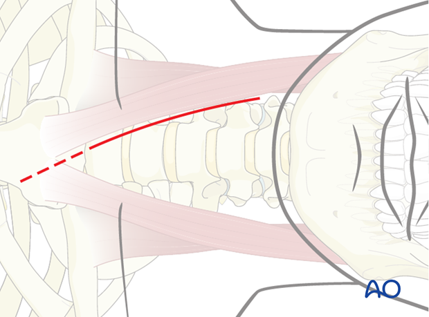
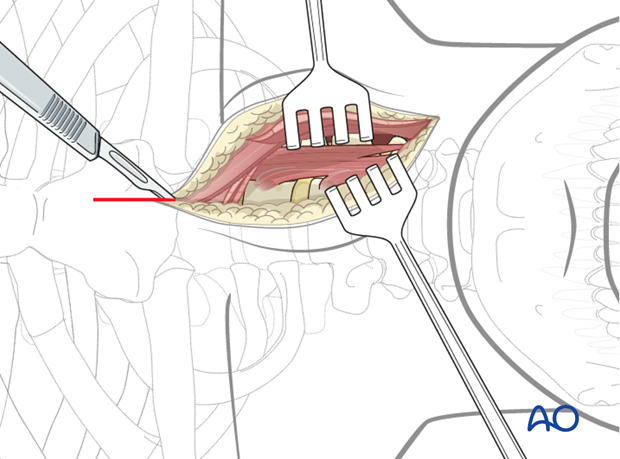
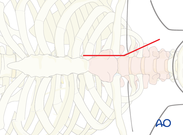
The incision is starts along the medial border of the sternomastoid muscle down to the sternal notch.
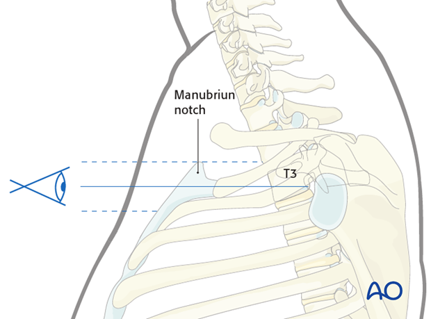
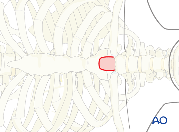
One may estimate how caudally the cervicothoracic junction can be accessed by viewing the position of the sternum relative the cervicothoracic junction on lateral radiograph or sagittal CT image.
If looking through the manubrium notch (in red) you can see the upper thoracic, then a common lower cervical would be enough, as in the T3 example.
If you only can see the lower cervical an extended approach would be recommended.
A left sided approach places the thoracic duct at risk while a right sided approach may be more likely to injure the recurrent laryngeal nerve.
3. Dissection
Platysma muscle is transected in line with the skin incision.
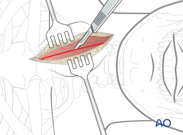
The deep cervical fascia is identified and divided along the anteromedial border of the sternocleidomastoid muscle.
It is beneficial to sufficiently release the superficial fascia as this greatly facilitates exposure of the deeper anatomic layers.
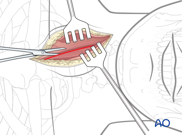
The omohyoid muscle crosses the surgical field at the level of C6 and can be transected to improve exposure.
Division of sternohyoid and sternothyroid muscles will facilitate the extensile exposure.
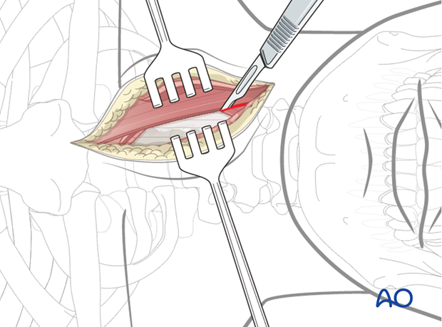
The carotid pulse is palpated and the dissection is directed medial to the carotid sheath.
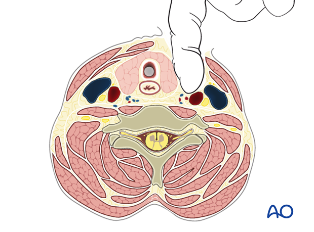
A finger is then used to dissect bluntly between the carotid sheath laterally and trachea and esophagus medially down to the prevertebral fascia.
Injuries to the esophagus and carotid artery can be associated with fractures or occur as a complication of the approach. Approach related complications can be avoided by using blunt dissection.
Esophageal injuries are serious and potentially lethal complications. The best results are achieved with immediate recognition and repair. Consultation with thoracic or ENT surgeons should be obtained.
Note: In case of carotid artery injury direct pressure should be applied and vascular surgery consultation requested urgently.
The incision is continued along the proximal third of the sternum.
Depending on the exposure needed, a manubriotomy may be sufficient to provide sufficient access.
If a more extensive approach is needed, a sternoclavicular approach may be used (as described below).
The sternoclavicular joint is opened. At this point, the medial end of approximately 3 cm of the clavicle may be resected to facilitate access.
The resected clavicle can be used as a strut graft.
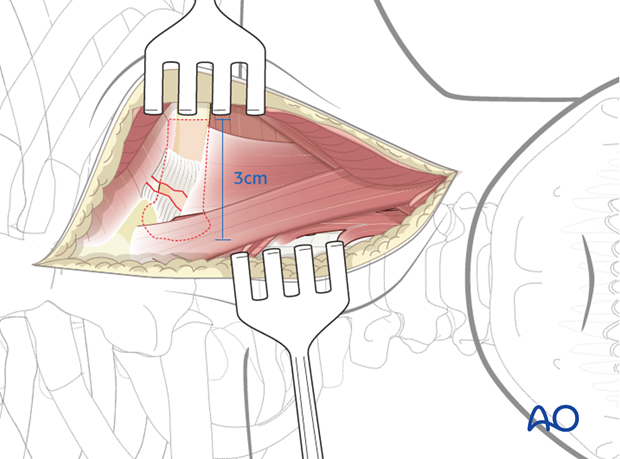
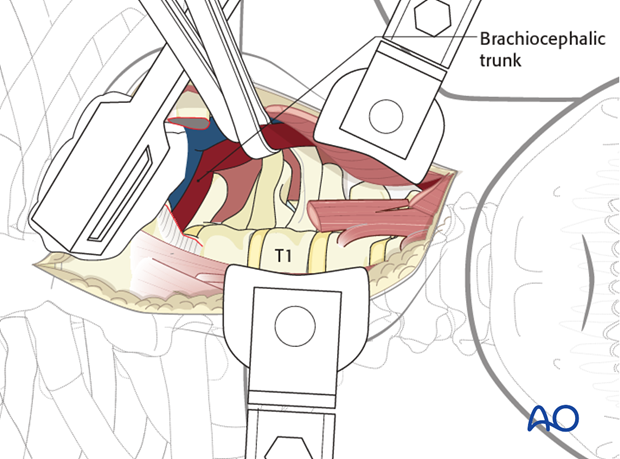
Blunt dissection is made down to the brachiocephalic trunk which is mobilized as needed.
The lung is protected using a sponge if needed.

The prevertebral fascia is cut longitudinally allowing direct visualization of the vertebra and the longus coli muscle.
The level is verified with fluoroscopy.
The sternothyroid, sternohyoid, and omohyoid muscles may be retracted or transected when necessary for a better exposure.
4. Closure
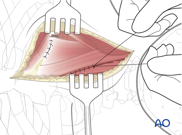
The split muscles are approximated and the transected sutured.
The platysma muscle is sutured followed by a subcutaneous and skin closure. Skin closure can be performed with a subcuticular suture to enhance cosmesis.
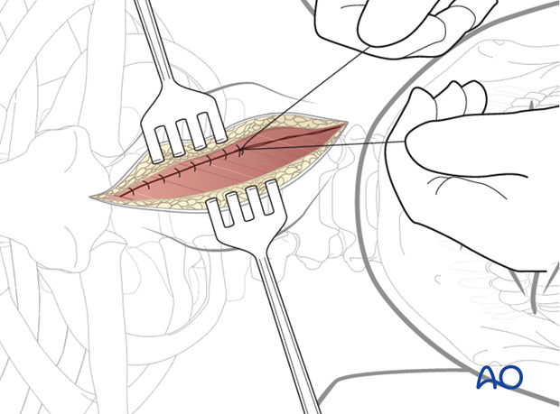
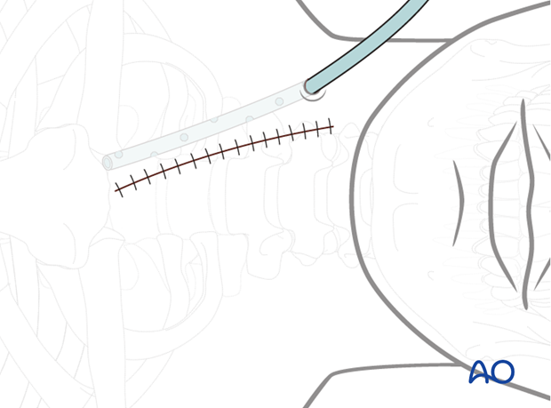
A wound drain is inserted through a separate stab incision.
