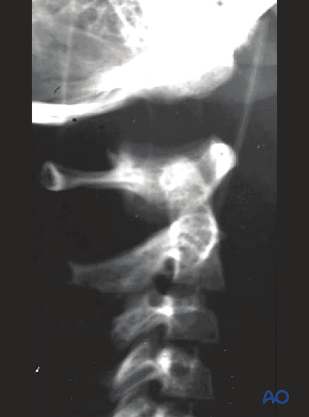
Highlights of Occipitocervical trauma
Introduction
Publication of second edition (May 2024)
On behalf of the AO Spine Trauma Knowledge Forum the Occipitocervical trauma module has been updated to show the new upper cervical injuries classification. This work was overseen by Cumhur Öner (the Netherlands). Luiz Vialle (Brazil) was the editor.
This module is based on current clinical principles, practices, and available evidence, and supports spine surgeons’ day-to-day treatment, planning, learning, and teaching.
Visit the module entry page here.

Highlights
The classification system provides surgeons from different institutions with a common language to discuss various injuries. It provides consistency in injury diagnosis and treatment.
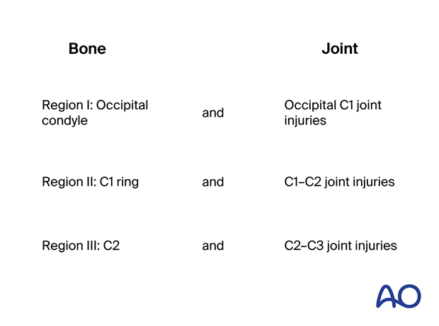
Unlike the subaxial, thoracolumbar, and sacral classifications, which have a uniform vertebral anatomy, the upper cervical spine has three distinct segments, with typical bones and joints. For this reason, this upper cervical area has been divided into three regions, as illustrated.
Each region involves a bone and its lower joint. In this way, it is possible to apply the morphological concepts already described in the A, B, C system to each one of the regions.
The new module details treatments tailored to different occipitocervical injuries. Additionally, the module offers injury definitions, treatment indications, guides to patient positioning, and approaches.
The module addresses:
- Clinical evaluation
- Neurological evaluation
- Radiological evaluation (XR, CT, MRI)
A wide range of basic techniques are also described.

Content
Table of contents
Region I: Occipital condyle and craniocervical junctionType A: Isolated bony injury (condyle):
- Nonoperative treatment (collars)
Type B: Nondisplaced ligamentous injury (craniocervical):
- Halo vest
- Occipitocervical fusion
Type C: Any injury with displacement:
- Occipitocervical fusion
Type A: Isolated bony injury:
- Nonoperative treatment (collars)
- Halo vest
Type B: Nondisplaced ligamentous injury:
- Halo vest
- Posterior C1–C2 fusion
- C1 lateral mass osteosynthesis
Type C: Translation injury of the C1–C2 joint:
- Halo vest
- Posterior C1–C2 fusion
- Occipitocervical fusion
Type A: Isolated bony injury of C2:
- Nonoperative treatment (collars)
- Halo vest
- Anterior odontoid screw
- Posterior C1–C2 fusion
Type B: Nondisplaced ligamentous injury:
- Nonoperative treatment (collars)
- Halo vest
- Posterior C2–C3 fusion
- Direct osteosynthesis of the isthmus
- Anterior C2–C3 fusion
Type C: Translation injury:
- Anterior C2–C3 fusion
- Posterior C2–C3 fusion
- Posterior C1–C3 fusion
Additional material
Approaches:
- Anterior access to C1–T2
- Posterior access to C1–T2
Patient preparations:
- Prone position
- Supine position
Further reading:
- AO Spine upper cervical injuries classification system
- Clinical evaluation
- Neurological evaluation
- Radiological evaluation (XR, CT, MRI)
Basic techniques:
- Anterior C1–C2 transarticular screw insertion
- C1 lateral mass screw insertion
- C2 laminar screw insertion
- C2 pars screw insertion
- C2 pedicle screw insertion
- Lateral mass screw insertion (Magerl technique)
- Occipital condyle screw insertion
- Pedicle screw insertion in the cervical spine
- Transarticular screw insertion


