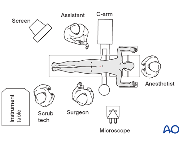
Prone position for approaches to L1–L5
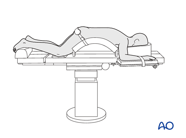
1. Patient positioning
The patient is positioned on a Wilson frame on a radiolucent table.
- The head is placed in a padded face mask to avoid pressure on the eyes and have the endotracheal tube free with the neck in a neutral position.
- The abdomen should hang free to avoid high intraabdominal pressure and subsequent venous pressure causing excessive bleeding of the spine.
- The arms/shoulders should rest comfortably in a 90° position of the shoulder and elbow. Ensure the wrists and elbows are adequately padded to prevent a peripheral neuropathy.
- Adequate padding must be provided to elbows and knees to avoid pressure sores.
- A small bump or blanket roll should be placed under the legs to flex the knees slightly.
Positioning should attempt to maintain/increase lumbar kyphosis, which opens up the interlaminar and foraminal spaces.
Pearl: When performing fusion surgery, the frame's kyphosis must be removed in order to achieve a lumbar lordosis before adding the rods. If this is not done, then post-surgical flatback may occur.
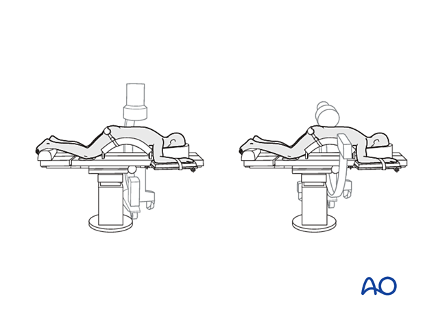
It must be possible to obtain radiographic images in both AP and lateral planes at all times.
2. Anesthesia
General anesthesia with endotracheal intubation is usually performed.

3. Preoperative antibiotics
Antibiotics should be administered prior to incision and at two-hour intervals during the procedure.
A cephalosporin antibiotic with good Gram-positive coverage is generally recommended.
Patients with penicillin allergies should receive vancomycin or clindamycin.
4. Neuromonitoring
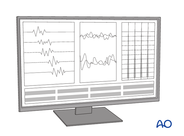
Neuromonitoring is optional for most decompressive surgeries and typically includes free running and triggered EMGs. In cases where patients also have cervical or thoracic stenosis, SSEP and MEP monitoring should be considered to ensure adequate spinal cord evaluation during the surgery.
5. Fluoroscopy
The incision can be planned based on AP and lateral fluoroscopy. Alternatively, intraoperative CT with three-dimensional navigation can be used.
OR setup for endoscopic procedures
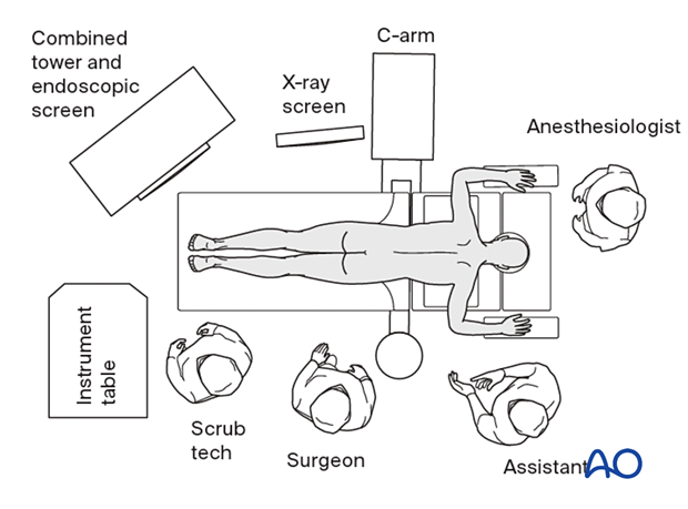
The endoscopic imaging screen should be placed at the foot of the bed on the opposite side of the surgeon. The fluoroscopy screen should be placed next to this on the opposite side of the surgeon.
OR setup for microscopic procedures
If a microscope is used, it should be placed on the surgeon’s side and opposite the image intensifier portion of the C-arm fluoroscope.
