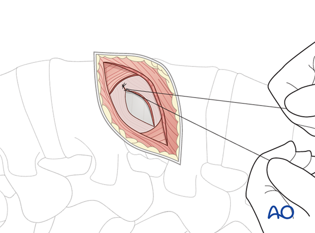
Minimally invasive transpsoas approach (L2–L4)
1. Portal site
The exact portal site will depend on the level of the disc disease. The disc space L5–S1 cannot be reached with this approach.
For minimally invasive surgery, specific instruments and implants are needed. A self-retaining retractor system is necessary to allow the exposure of the spine.
It is necessary to confirm the correct level of the approach with fluoroscopy.
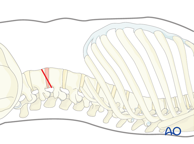
2. Application
The lateral transpsoas is ideal for cage insertion. It is a retroperitoneal minimally invasive approach.
3. Preparation
Under fluoroscopic control, the disc space is marked on the skin.
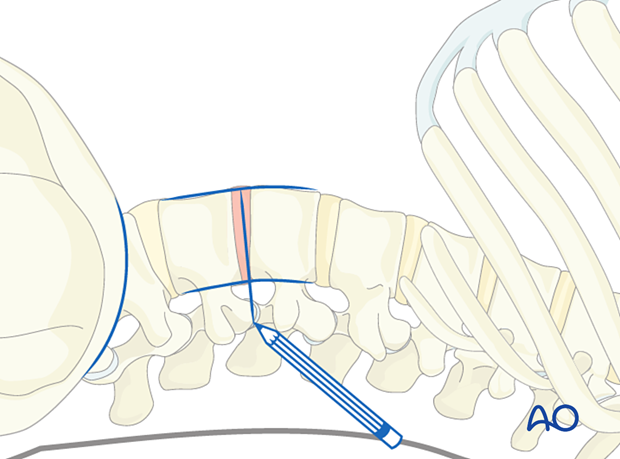
The incision line should be made exactly in the direction of the target. The length of the incision is usually approximately 3–4 cm (depending on how many levels will be treated).
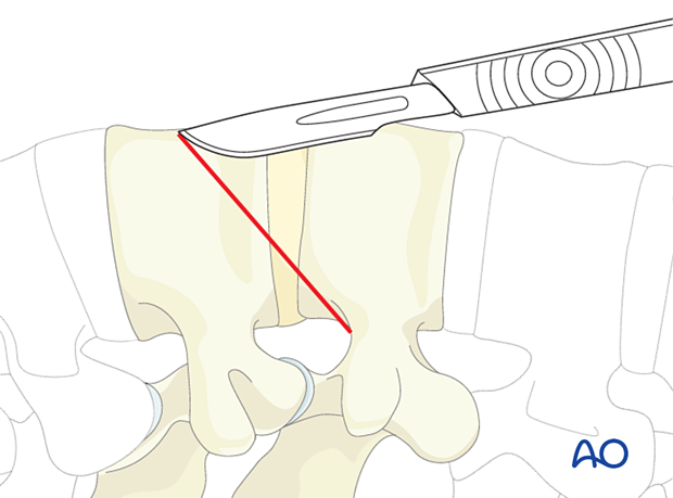
4. Exposure
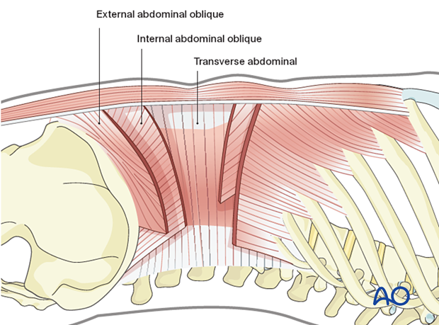
There are three abdominal wall muscles. The first layer is the external abdominal oblique muscle, the second layer is the internal abdominal oblique muscle, and the third layer is the transverse abdominal muscle.
The subcutaneous tissue and the fascia of the external abdominal oblique muscle are dissected.
The first muscle layer is split with scissors and a finger and retracted.
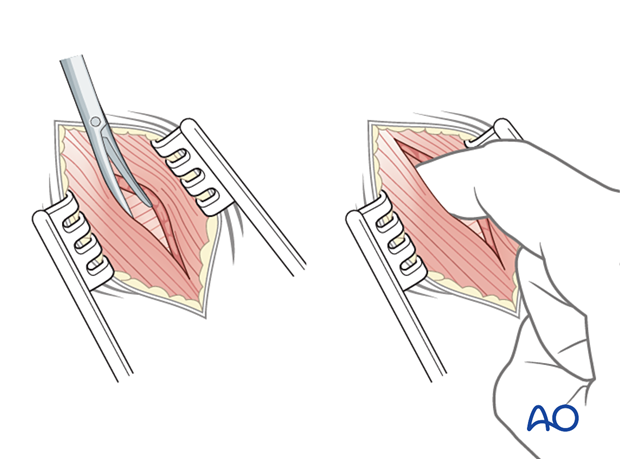
The second layer is split and retracted (the orientation of the fibers of the second layer is roughly perpendicular to the first layer). The retractors should be positioned parallel to the orientation of the fibers.
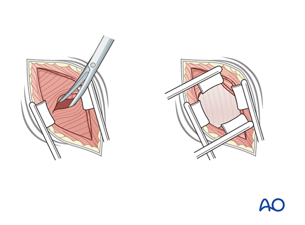
The fascia of the transverse abdominal muscle is opened with caution to avoid injury to the peritoneum, which lies in the abdominal cavity. A finger should be used at this point to identify the edge of the disc space through palpation.
Retroperitoneal fat typically falls away from the abdominal wall.

To facilitate dissection, a finger is used to detach the psoas muscle from the peritoneum. The finger should always follow the fibers of the psoas muscle without wandering anteriorly or posteriorly.
The retroperitoneal fat is a good landmark for detecting the retroperitoneal space. It may be more difficult to identify this fat in very thin patients.
Fat is stripped off the abdominal wall from dorsal to ventral, starting at the incision using finger dissection.
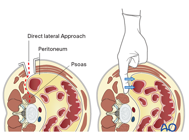
Pitfall: Injured peritoneum
If the peritoneum is injured, intraperitoneal organs can be affected, causing abdominal issues.
If the peritoneum is violated, it is recommended that it should be repaired immediately with an absorbable suture and a blunt tapered needle.
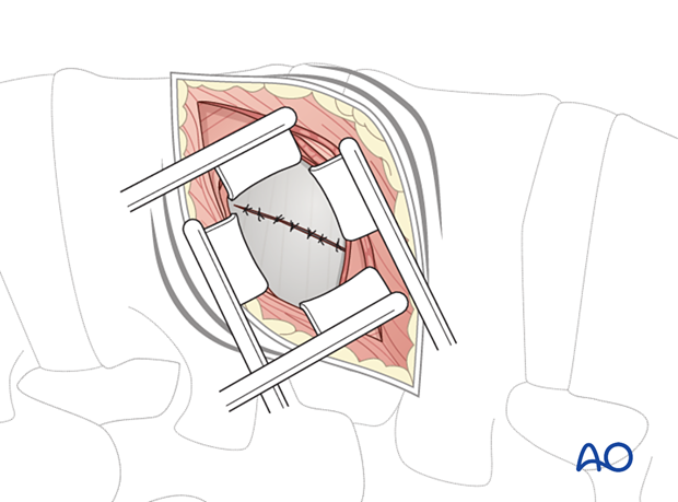
The psoas muscle is exposed to facilitate posterior retraction.
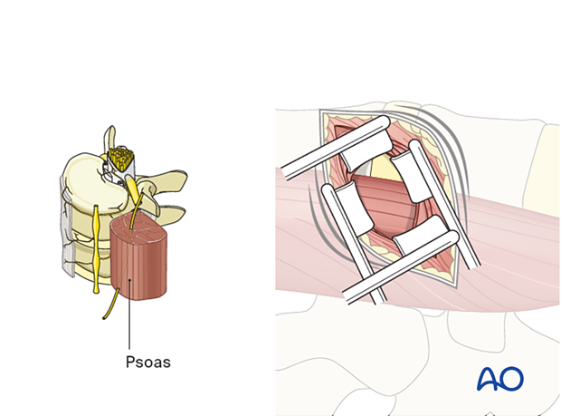
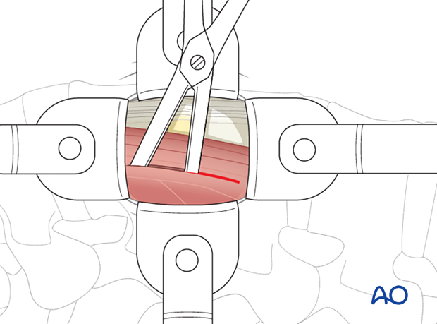
Following x-ray confirmation of the correct level, the psoas muscle is gently split with scissors or a finger for a length of approximately 3–4 centimeters.
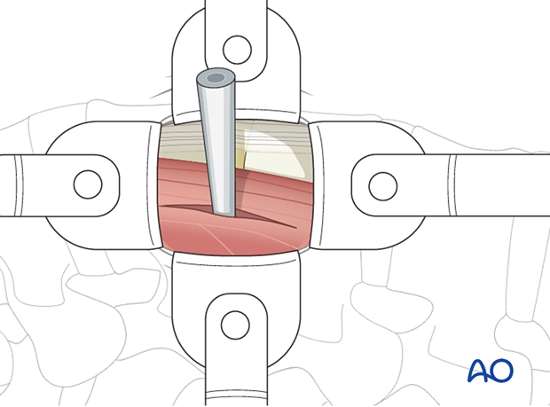
A tubular localizer is positioned through the muscle over the edge of the disc space. This position should be confirmed with AP and lateral views.
5. Closure
A standard closure of the fascia is recommended. The wound is then closed in layers.
