Primary repair with pedicle screw and rod
1. Comment on approach
Fluoroscopy is used to confirm the correct level of L5 and S1.
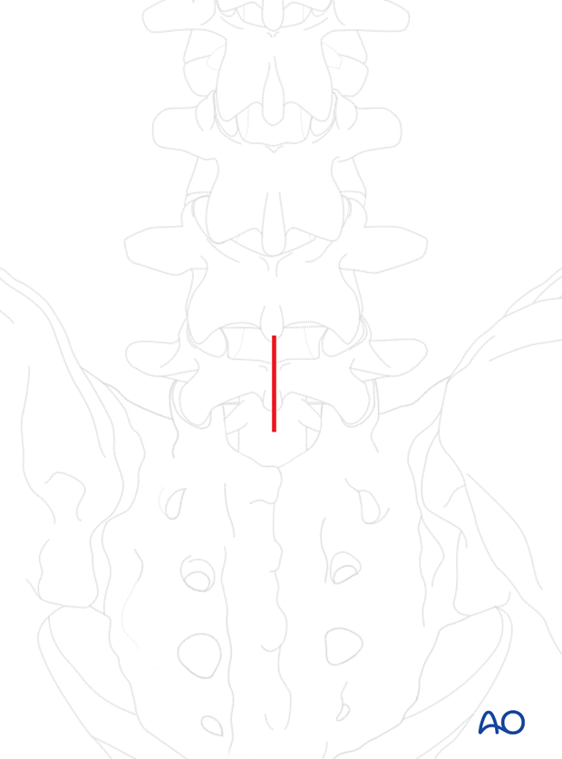
A midline skin incision is performed with the patient in prone position to access the L5-S1 joint.
The subperiosteal dissection is taken down onto the lamina of L5 exposing the L5-S1 joint
Care must be taken not to violate neither the L5-S1 joint nor the L4-L5 joint.
Dissection is carried laterally onto the transverse process of L5.
2. Instrumentation
Pedicle screw insertion
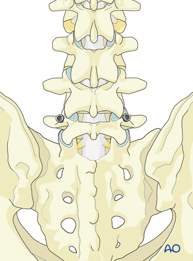
Pedicle screws are inserted bilaterally into L5 taking care not to breach the L4-L5 facet.
Hook placement
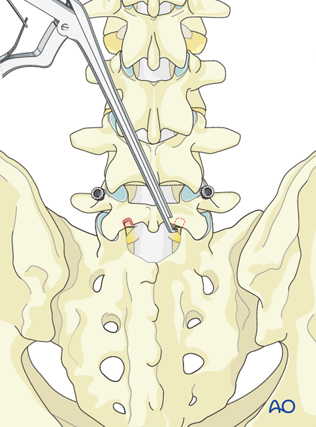
A small notch is created on the inferior aspect of the L5 lamina in preparation for a sublaminar hook.
3. Decortication
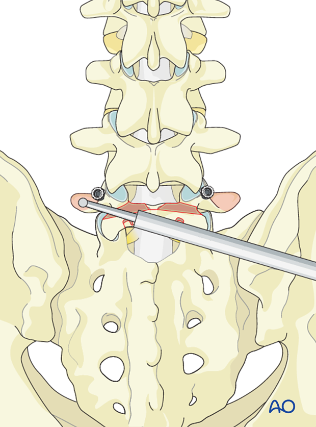
Using a high speed burr the defect in the pars articularis must be decorticated and the pseudoarthrosis excised down to bleeding bone.
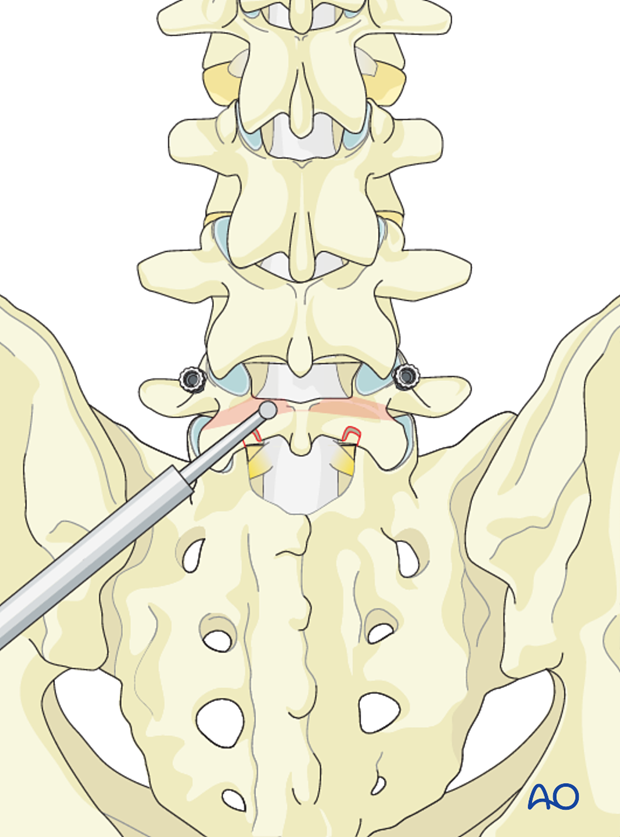
Decortication is also taken to the transverse process of L5 along the lamina of L5.
4. Bone grafting
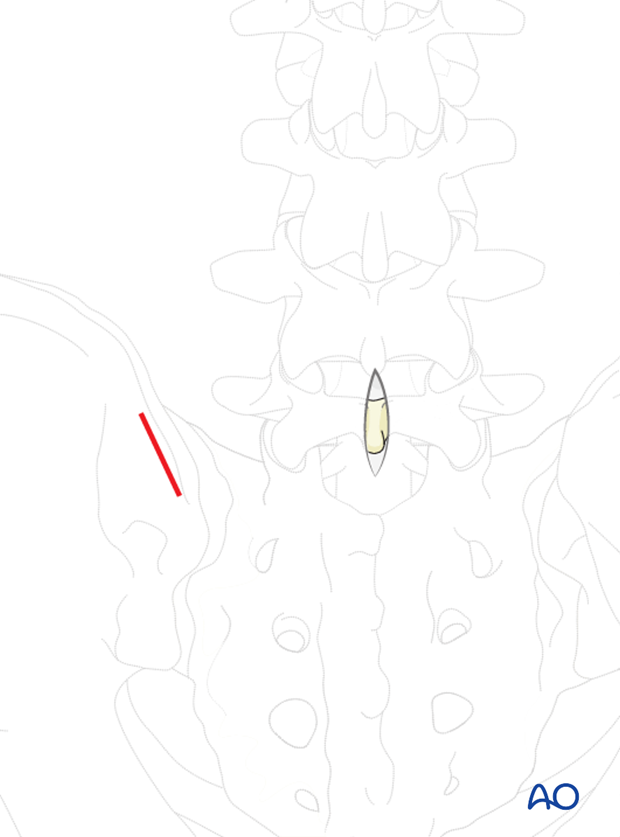
A separate incision is made over the posterior superior iliac crest.
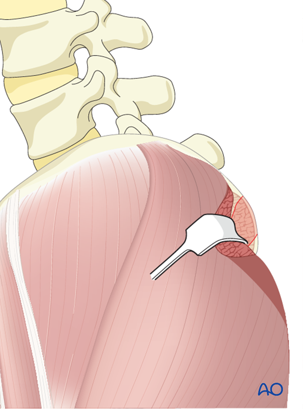
Muscle attached to the outer iliac crest is reflected in a blunt fashion exposing the outer iliac crest.
Using an osteotome or a gouge, corticocancellous strips are harvested in 4-5 cm length and 1 cm wide.
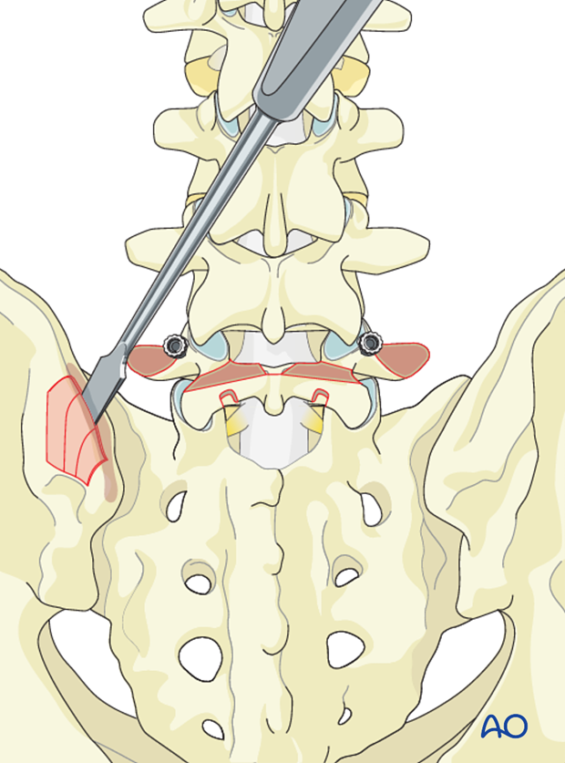
Smaller cancellous bone graft is also harvested from the iliac crest.
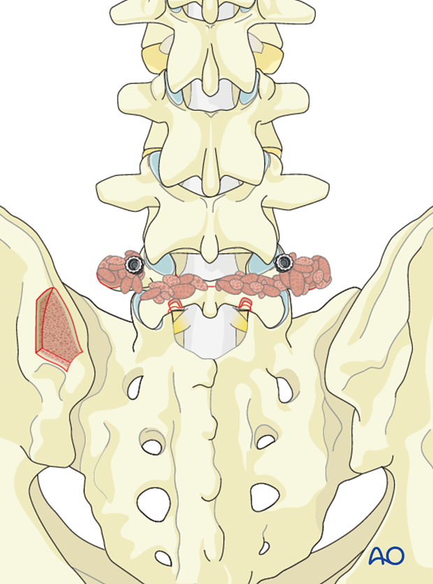
Bone graft must be impacted into the pars defect as well as the cortico cancellous strips are laid across the lamina and transverse process.
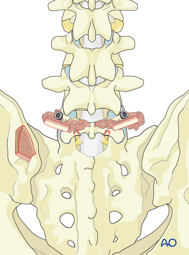
5. Rod insertion
Bilaterally, laminar hooks are inserted.
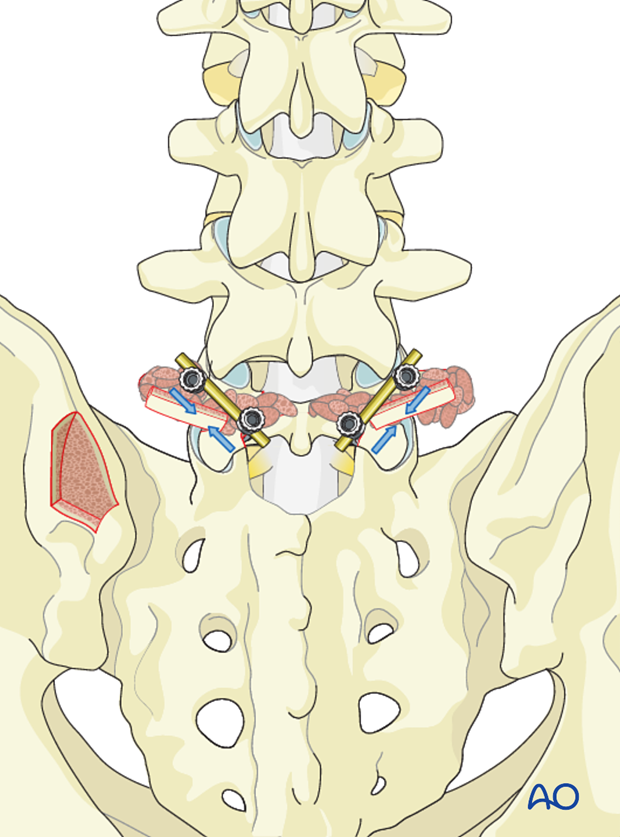
Short rods are connected to the laminar hooks and pedicle screws and compression is applied to close the pars defect.
6. Aftercare
Detailed postoperative neural assessment must be conducted, specifically looking at the integrity of the L5 nerve root as well as sacral nerves controlling bowel and bladder.
Patients with high grade spondylolisthesis that have been reduced are at high risk of postoperative foot drops secondary to neuro traction injury. To minimize such injuries patients are immediately placed in bed with knees and hips flexed. As long as no neuro injury can be identified the leg can be gradually extended after day two. If neuro injury can be identified, the period of flexion should be extended.
Patients are made to sit up in the bed on the first day after surgery. Bracing is optional. Patients with intact neurological status are made to stand and walk on the second day after surgery. Patients can be discharged when medically stable or sent to a rehabilitation center if further care is necessary. This depends on the comfort levels and presence of other associated injuries.
Patients are generally followed with periodical x-rays at 6 weeks, 3 months, 6 months, and 1 year looking for spinal fusion.
Patients with a diagnosis of dysplastic spondylolisthesis run a higher risk of cauda equina and require closer monitoring of their neurological status during and after surgery.


