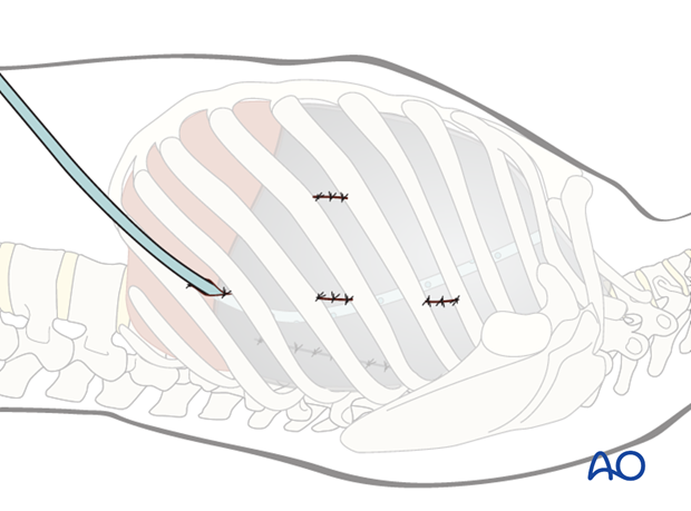
Anterior thoracoscopic release
1. Portals
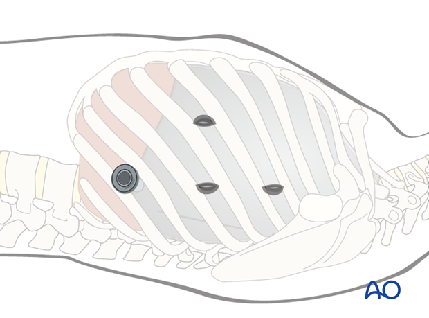
The exact portal site will depend on the level of the apex of the spinal curvature and the degree of vertebral orientation.
In principle, the instrument portals should be in line with the anterolateral aspect of the spine to allow removal of the disk and the anterior longitudinal ligament without risk of entering the spinal canal. Typically this is in the region of the anterior axillary line.
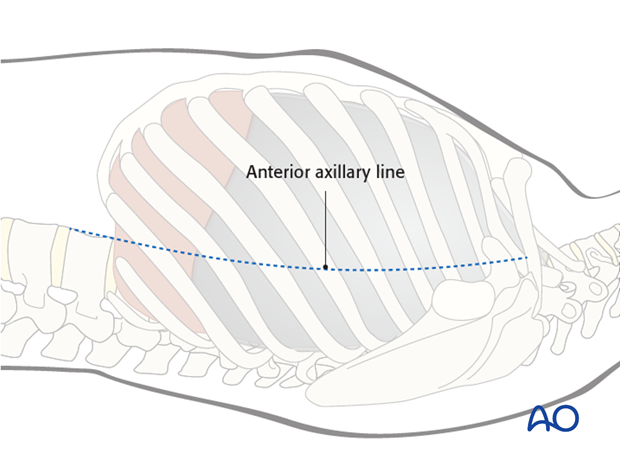
The viewing portal is prepared first and introduced from inferior making sure to be above the insertion of the diaphragm at approximately T10.
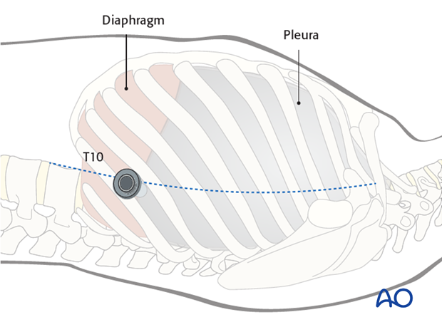
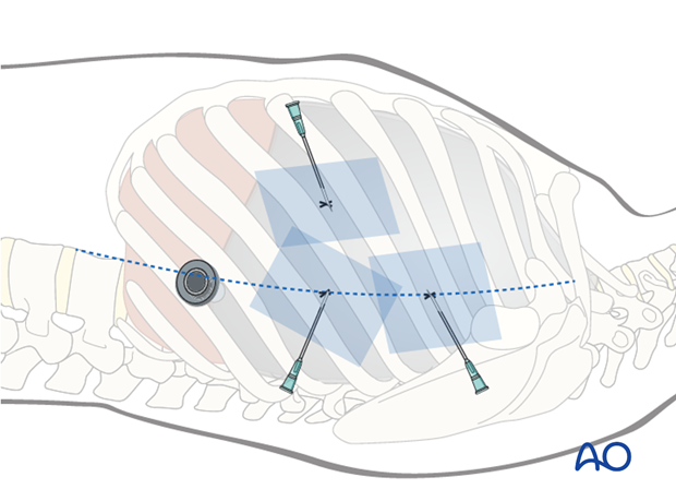
A needle is then introduced through any further planned portal sites and the correct entry points into the chest cavity verified under direct vision and/or using image intensification.
2. Instruments for thoracoscopic approaches
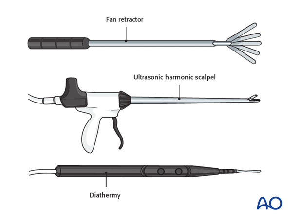
Specialized long instruments are needed for dissection. As an example a fan retractor can be used to carefully hold long or diaphragm tissue away from the vertebrae.
An ultrasonic harmonic scalpel is a very useful tool for both cutting and coagulating tissues if available. An alternative would be a long hook shaped diathermy tip.
3. 1st portal
A 10 mm skin incision is made in the lower part of the chest cavity between the seventh to tenth intercostal spaces in the anterior axillary line.
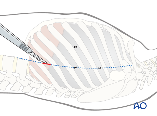
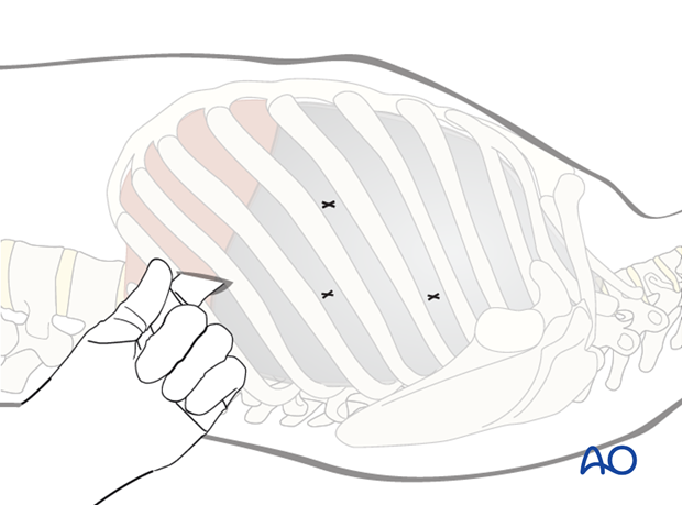
The incision is deepened by sharp dissection to the pleural cavity above the rib to avoid injury to the intercostal vessel and a finger is introduced into the chest cavity to check for adhesions.
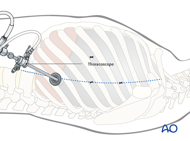
A blunt trocar is introduced into the chest space followed by the thoracoscope. This is used to examine the chest cavity for adhesions or anatomical variations prior to preparation of any further portals.
4. 2nd portal
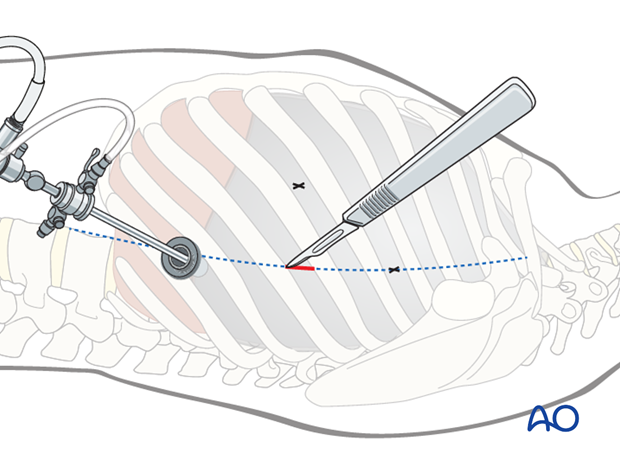
The second portal is located directly opposite to the apical disk in the anterior axillary line.
After verification of the entry point under direct vision, the incision is made followed by sharp dissection to the pleural cavity.
This is the working portal mainly used for performing dissections and the diskectomies.
5. 3rd portal
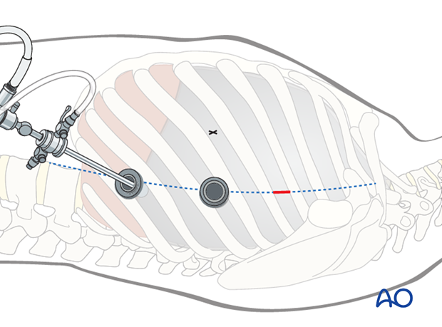
As the disk spaces are slanting away from the apex, each instrument portal can typically, at most, only approach 2 -3 disk spaces. The third portal is therefore prepared cranial to the second portal, to allow access to the higher disc spaces and to retract the lung.
The portal is then prepared in the same way as the 2nd portal.
6. Additional portals
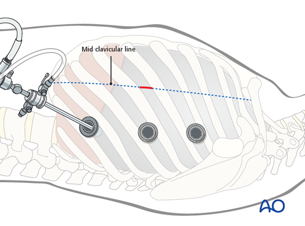
Additional portals can be made in line with the previous ones to allow access to additional disc spaces.
On occasions, an additional anterior portal can be made directly opposite to the apical disk in the mid clavicular line for lung retraction and to aid dissection. Care should be taken to avoid the internal mammary artery, visible from inside the chest wall.
The portal is prepared in the same way as the 2nd portal.
7. Further dissection
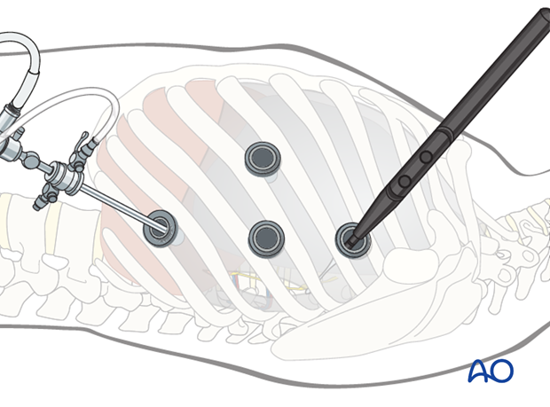
The parietal pleura over the appropriate disc spaces are divided longitudinally. The segmental vessels running along the vertebral bodies are identified. For anterior discectomies, these can be spared.
8. Closure
The viewing portal should be removed last to allow a direct inspection of the pleural cavity. The anesthesiologist should be asked to fully inflate the lung and this can be checked by the thoracoscope.
A chest drain is then inserted via this portal, lying on the posterior chest wall and extending all the way up to T2.
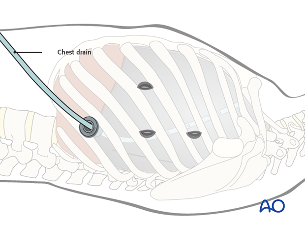
The portals are then closed in layers.
