Hook insertion for management of adolescent idiopathic scoliosis
1. Cranially pointing hooks
Pedicle hooks
Pedicle hooks can be inserted in a cephalad fashion typically between T1 and T10.
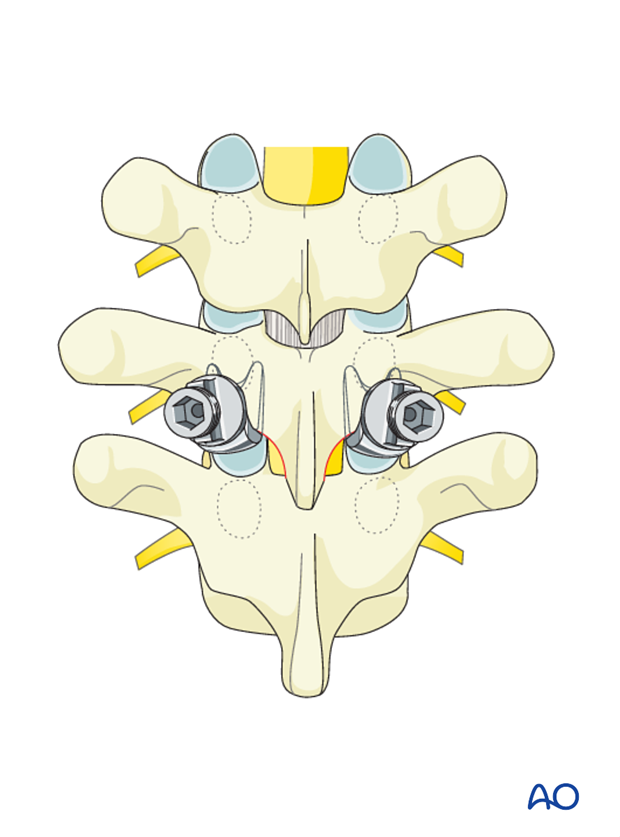
To ease insertion of the pedicle finder in a safe manner, a partial inferior facet excision should be performed.
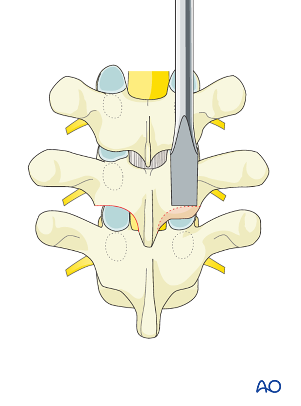
The pedicle finder is carefully positioned under the inferior facet and above the superior fact joint and directed along the vertical axis of the vertebra until it reaches the inferior pedicle pole.
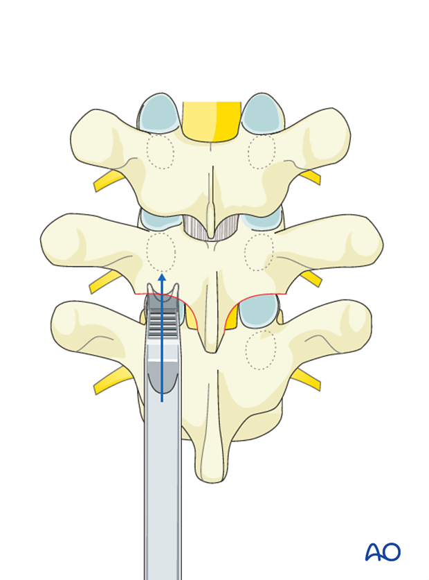
The hook is inserted directly towards the inferior pedicle while taking care not to direct the hook medial nor ventral towards the spinal canal. Ideally the hook should sit in a very stable position within the residual facet joint firmly against the pedicle.
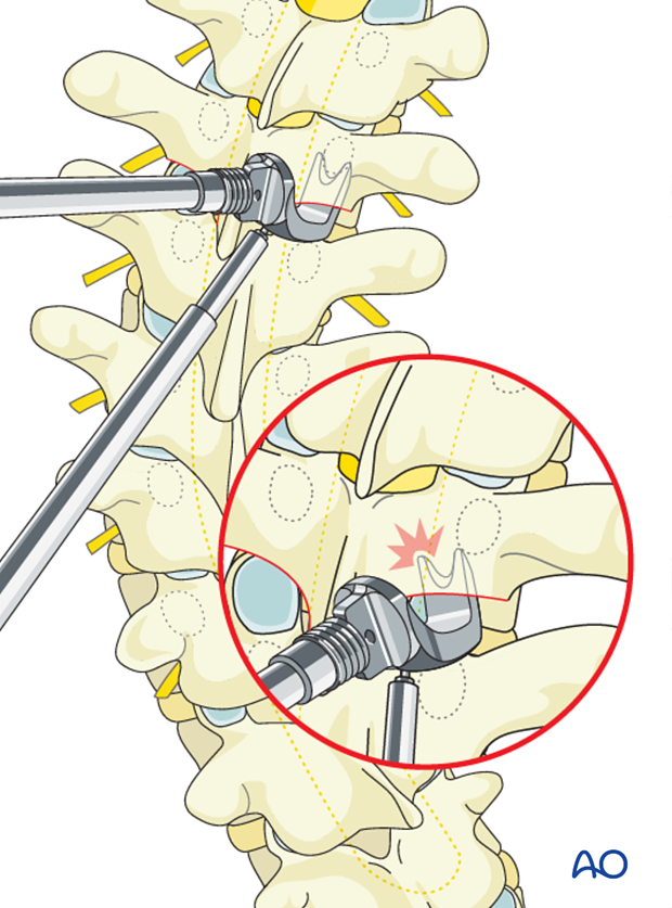
Sublaminar hooks
Cranially pointing sublaminar hooks can be used anywhere within the thoracic or lumbar spine.
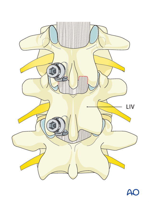
A laminar developer is utilized to develop an interval between the inferior edge of the inferior lamina and the ligamentum flavum.
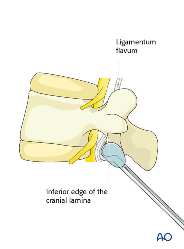
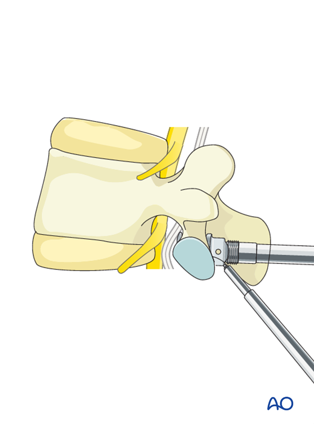
The hook is inserted in the interval developed by the laminar developer.
Correct insertion will ensure that the spinal canal is not entered and the base of the hook is resting snugly against the laminar surface.
2. Caudally pointing hooks
Transverse process hooks
Transverse process hooks can be used in the thoracic spine typically between T1 – T10.
The transverse process finder is used to expose the cranial edge of the transverse processes superperiosteally ensuring firm purchase along the superior edge of the transverse process.
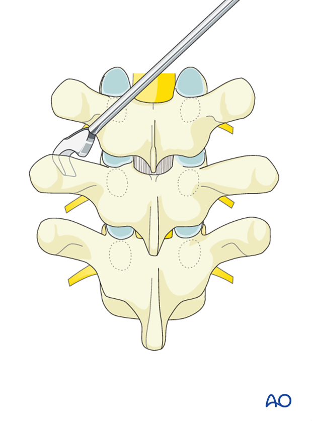
A wide blade hook is placed firmly against the superior transverse process ridge at the midpoint of the mediolateral portion of the transverse process.
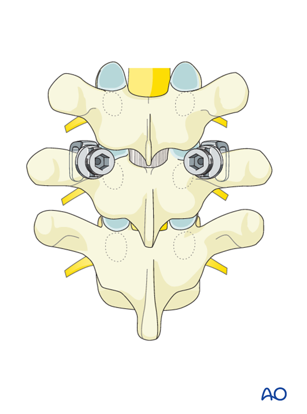
Supralaminar hooks
Caudally pointing supralaminar hooks can be used at any level of the thoracic and lumbar spine.
The ligamentum flavum is removed from the cranial edge of the lamina unilaterally at the site of hook placement. Occasionally laminar bone removal may be required to create enough space to allow the hook blade to safely enter the spinal canal. Also ensure that the dura is not adherent to the undersurface of the lamina.
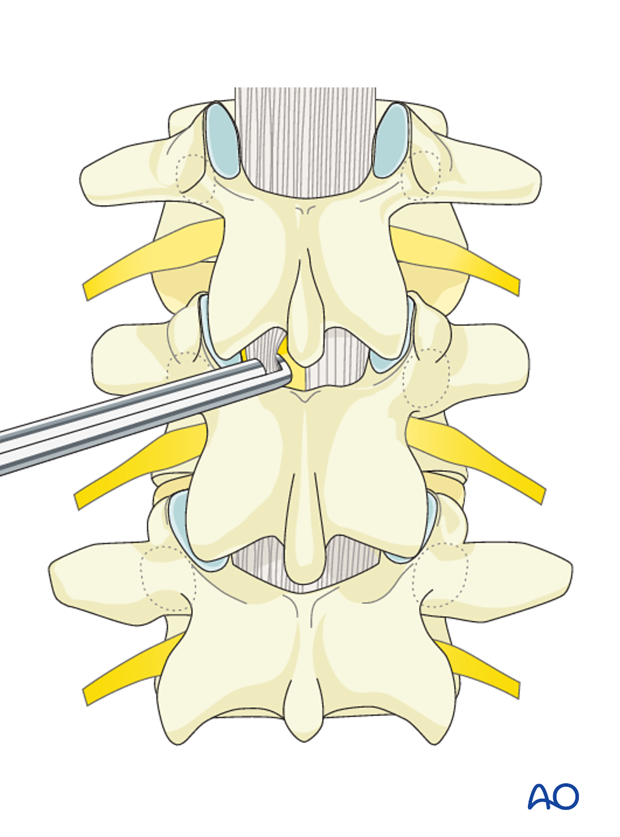
The hook blade is carefully placed on top of the dura under the surface of the lamina. Great care must be taken to ensure that the body of the hook hugs the laminar surface both during and after insertion to avoid ventral encroachment of the spinal canal by the hook blade.
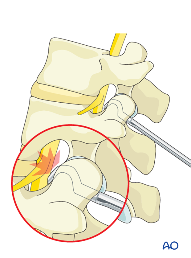
Laminar hooks can be inserted on consecutive levels of the spine when required.


