Patient preoperative evaluation
1. Introduction
Periprosthetic fractures are difficult to treat. Successful treatment requires planning and preparation. Information about the original arthroplasty procedure and implant is needed in addition to the usual information about the patient and the fracture.
2. Patient evaluation
Patients with periprosthetic fractures often present additional challenges, including the following comorbidities:
- Frailty
- Dementia
- Osteoporosis
- Sarcopenia
- Prior or current periprosthetic infection
Diagnostic workup needs to include an assessment of these factors.
3. Postoperative planning
It is important to verify whether the patient can cope with activities of daily living using one arm. If not, supportive patient care must be planned accordingly.
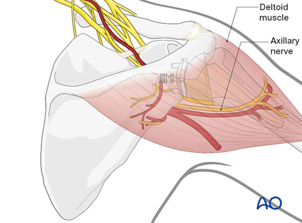
A general neurovascular examination, and a thorough assessment of the axillary nerve and the deltoid muscle innervation are important for decision making in periprosthetic shoulder fractures.

4. Prosthesis evaluation
Ideally, details of the original arthroplasty operation should be checked. It is useful to know the manufacturer and model of the prosthesis, the date of insertion, details of any perioperative complications, and information regarding prosthesis function.
5. Fracture evaluation
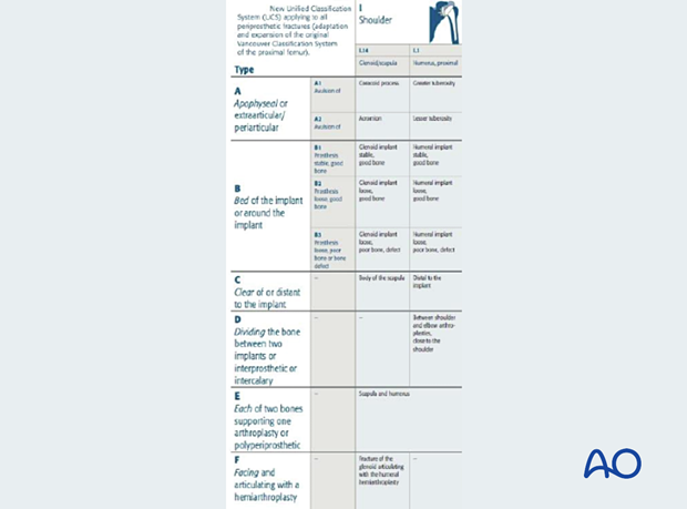
To evaluate and classify a periprosthetic fracture, the surgeon needs to assess not only the fracture location and configuration but also the presence of prosthetic loosening and the quality of bone stock. The Unified Classification System for Periprosthetic Fractures (UCPF) is a useful model that helps with this evaluation.
Diagnostic imaging needs to be interpreted in the context of clinical history and examination to enable a full evaluation of the prosthesis as well as the fracture. For example, regardless of imaging findings, indicators of possible periprosthetic loosening include:
- Preceding pain
- Deteriorating function
- Fracture with no history of trauma
- Previous or current infection
It is important to exclude prosthetic joint infection and septic loosening. Clues to the presence of infection can be obtained on history. They may include the following:
- Previous treatment for infection
- Postoperative wound problems (eg, prolonged serous ooze)
- Preceding pain
If an infection is suspected, diagnostic workup should include serologic workup, aspiration of the joint for cell count, microbiology, and deep tissue samples from the prosthesis interface taken at surgery. This may need to be done as a separate procedure before surgery for the periprosthetic fracture, either by open or arthroscopic surgery, especially if revision arthroplasty is planned.

6. Diagnostic imaging
It is important to establish if the prosthetic component is loose, because this determines whether internal fixation or revision surgery is the optimal treatment. Plain x-rays are useful, especially if previous images are available: this facilitates a comparison of the position and fixation of the components over time.
X-ray signs include:
- Periprosthetic lucency
- Prosthetic subsidence
- Changes in alignment and osteolysis
CT scans may show detail of osteolysis and occult fractures.
Advanced MRI techniques can give more detail of the bone-implant interface and soft tissues.


