Avulsion of the coracoid process
General considerations
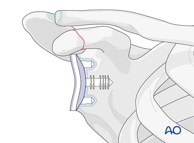
Fractures of the coracoid process are classified by UCPF as I.14-A1.

Fractures of the coracoid process are rare.
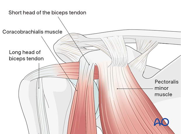
The coracoid process is the key to scapular control. The following muscles are attached to the coracoid process:
- Subclavius
- Pectoralis minor
- Short head of biceps
- Coracobrachialis
The following ligaments are attached to the coracoid process:
- Medial coracoclavicular ligament
- Trapezoid and conoid ligaments
- Coracoacromial ligament
- Coracoglenoid ligament
- Coracohumeral ligament
These structures, together with the acromioclavicular joint and clavicle, comprise the lateral scapular suspension system (LSSS). Detailed information about the LSSS is provided here.

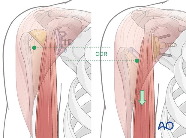
In reverse total shoulder replacement, the humeral center of rotation is usually more distal and medial than in anatomic total shoulder replacement. As a result, the tension in the tendons of the coracobrachialis and the short head of biceps (the conjoint tendon) is increased.

Etiology
Traumatic fractures
Avulsion can be caused by forceful contraction of the biceps or the coracobrachialis.
Avulsion can also happen in the case of anterior dislocation of the shoulder.
Fractures of the scapular body can extend to the coracoid base; in which case the pull of the tendons can cause displacement.
Stress-induced fractures
The lengthening of the arm in reverse shoulder arthroplasty can cause fatigue/stress fractures of the coracoid process, particularly in osteopenic bone.
Clinical signs
- Anterior shoulder pain which worsens during shoulder flexion or elbow flexion against resistance
- Weakness of shoulder flexion, elbow flexion, and supination
- Tenderness at the front of the shoulder on deep palpation
Imaging
Plain x-rays, particularly the scapular Y view, can demonstrate the fracture, especially in displaced cases. CT is recommended in suspected fractures and to evaluate displacement. For fatigue fractures, nuclear imaging can be helpful.


