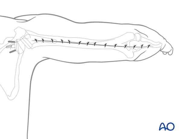
Posterior humeral approach
1. Introduction
This approach is most commonly used for periprosthetic fractures where the prosthesis is stable and is retained (B1 and C types) involving the distal half of the humerus. It can be extended for more proximal fractures after identification of the radial nerve.
2. Skin incision
Incise the skin, beginning at the tip of the olecranon.
The incision runs proximally in a straight line along the posterior midline of the arm. It crosses the trajectory of the radial nerve in the mid-humeral region and the axillary nerve proximally.

3. Superficial dissection
Incise the deep fascia in line with the skin incision.
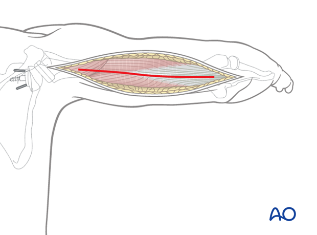
4. Deep dissection
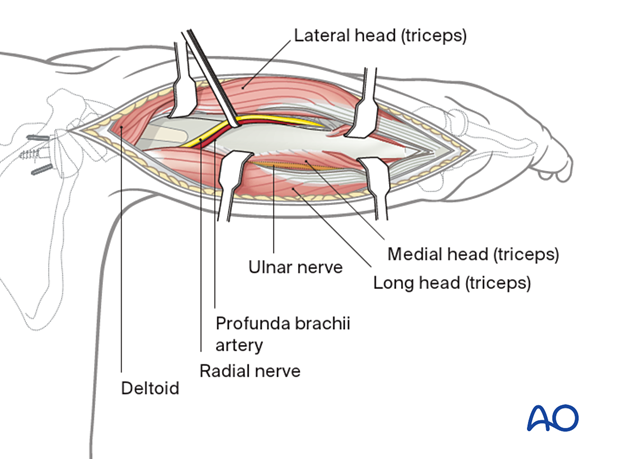
The interval between the lateral and long heads of the triceps is identified by palpation. The interval is developed from proximal to distal, noting the radial nerve and its branches beneath the triceps as it crosses the humerus.
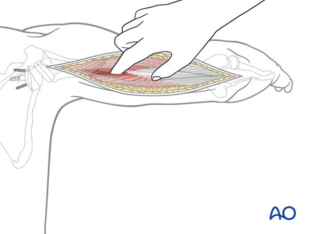
The radial nerve and accompanying profunda brachii artery are identified proximal to the medial head of the triceps in the spiral groove.
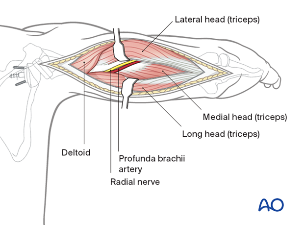
The triceps tendon and muscle are split distally by sharp dissection.
5. Extending the approach
Distal extension
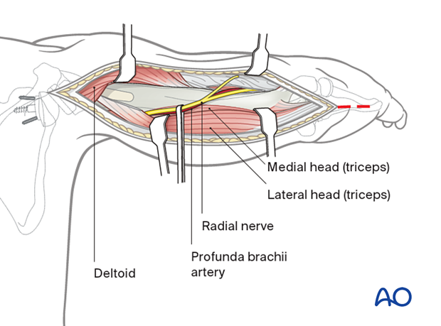
The triceps can be split to the level of the olecranon and continued into the forearm if necessary, as the Boyd-Thompson approach.
Proximal extension
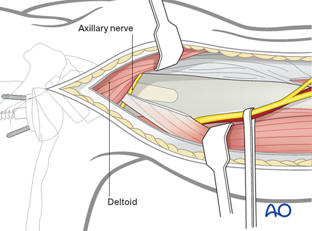
The posterior aspect of the proximal humerus can be exposed proximal to the radial nerve by retraction of the posterior border of the deltoid. Care must be taken not to injure the axillary nerve and its branch, the posterior superior cutaneous nerve of the arm.
6. Wound closure
The subcutaneous fascia and the skin are closed in layers.
