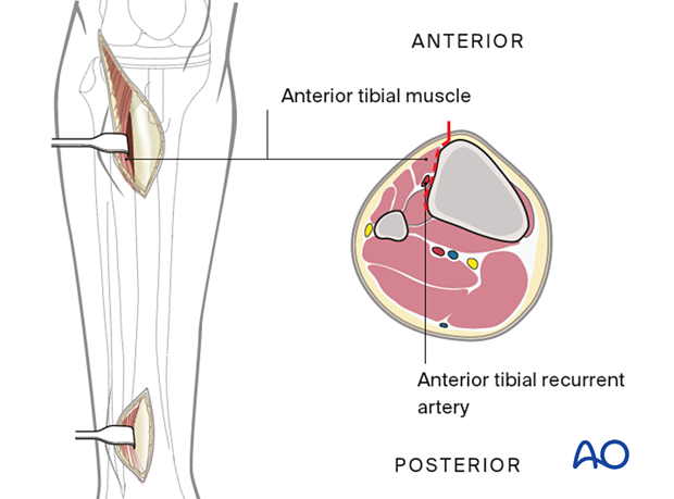
MIO Anterolateral approach to the proximal tibia
1. Indications
This approach is used for the fixation of proximal and mid-tibial periprosthetic fractures. The surgeon must balance the cost of a new incision over-utilizing the prior total knee incision. The skin between the two will be at risk, especially when the two surgeries are close in time to each other.
Distal fixation in the proximal third or midshaft can be achieved through stab incisions.
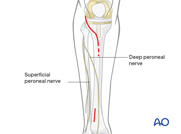
Over the distal third of the tibia, a short incision is advisable in this region to expose and retract the deep peroneal nerve and anterior tibial vessels, to avoid their being trapped by a submuscular plate, or injured by percutaneous screws.
2. Anatomy
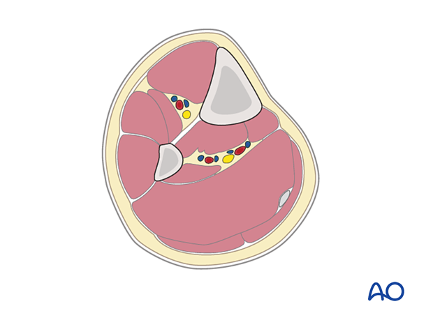
Triangular shape of the tibia
The lateral and posterior surfaces of the tibia are covered by muscle. The anteromedial surface has only a thin layer of subcutaneous tissue and skin. This surface provides less blood supply to the underlying bone.
Compartments
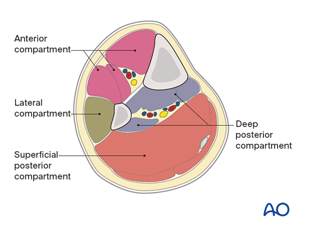
The lower leg has four compartments:
- Anterior
- Lateral
- Deep posterior
- Superficial posterior
The anterior compartment has three muscles and one main artery and nerve: Tibialis anterior, extensor hallucis longus, extensor digitorum longus; the anterior tibial artery, and deep peroneal nerve.
The lateral compartment has two muscles and one nerve. The muscles are the peroneus longus and brevis and the superficial peroneal nerve.
The deep posterior compartment has three muscles and two arteries and one nerve: The muscles are the tibialis posterior, the flexor hallucis longus, and the flexor digitorum longus. It also has the peroneal artery and the posterior tibial artery as well as the tibial nerve.
The superficial posterior compartment has just two muscles in it: the gastrocnemius and soleus muscles and the sural nerve.
3. Proximal skin incision
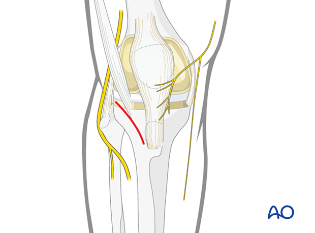
Identify Gerdy’s tubercle. Make a straight incision about 5cm in length starting posteriorly to Gerdy’s tubercle and running distally and anteriorly.
4. Distal skin incision
The incision in the distal third is made to prevent neurovascular injury from percutaneous screws. The anterior tibial vessels and deep peroneal nerve cross the anterolateral surface of the tibia approximately at the junction of its third and fourth quarter.
Stab incisions can be made in the proximal half of the tibia just lateral to the tibial crest.

5. Deep dissection
For the proximal wound, open the deep fascia starting at Gerdy’s tubercle and extend it distally. Expose the bone by releasing the anterior attachment of the tibialis anterior.
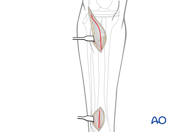
At the distal incision dissect the tibialis anterior tendon from the bone, preserving the paratenon.
Retract the tendon together with the deep peroneal nerve and the anterior tibial vessels to the lateral side.
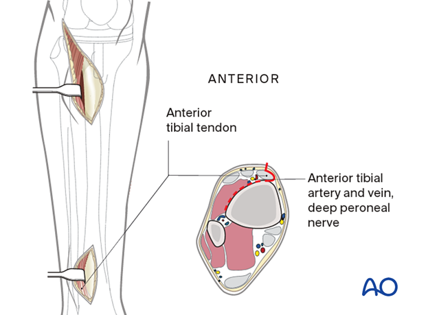
Deep dissection of stab incisions in the proximal half of the tibia can be performed by splitting the anterior compartment muscles using blunt dissection.
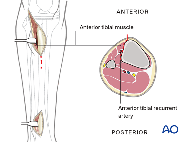
At the proximal incision, dissect the tibialis anterior muscle from the bone without stripping the periosteum.
Retract the tibialis anterior muscle laterally.
