Patient preparation in supine position
1. Introduction
The supine position is used for the treatment of most tibial shaft fractures.
2. Preoperative preparation
Read the additional material on preoperative preparation.
3. Anesthesia
Fracture manipulation and surgical stabilization are usually performed under general anesthesia.
Regional and local anesthetic blocks may be used to reduce postoperative pain.
Regional blocks should be avoided in patients at risk of developing compartment syndrome.
4. Prophylactic antibiotics
Antibiotics are administered according to local antibiotic policy and specific patient requirements.
5. Patient position
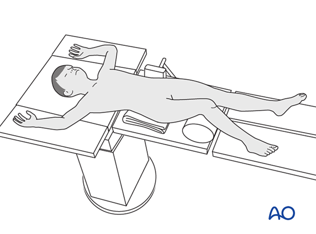
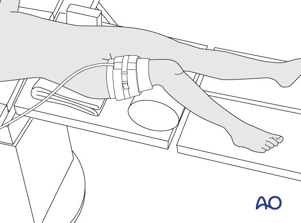
Place the patient supine on a radiolucent table.
A bolster or roll under the knee may be used to flex the knee.

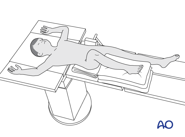
Alternatively, the leg may be elevated with a stack of towels.
This allows intraoperative AP and lateral imaging without manipulation of the extremity.
6. C-arm positioning
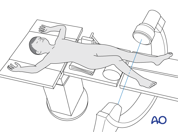
Position the C-arm so that AP and lateral views can be obtained, with the beam orientated from the uninjured side.
7. Tourniquet use
A tourniquet is usually not necessary but can reduce blood loss and improve visualization in selected cases.
The effect of ischemia/reperfusion in the presence of a compromised soft-tissue envelope must be considered.
The tourniquet width should be more than half the limb diameter and applied over a thin layer of padding.
The ischemic time should not exceed 120 minutes.
8. Skin preparation and draping
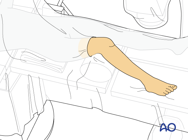
Prepare the lower leg, including the distal femur and foot.
Drape the limb to the mid-thigh or the level of the tourniquet.
Drape the C-arm in a conventional manner.
9. OR set-up
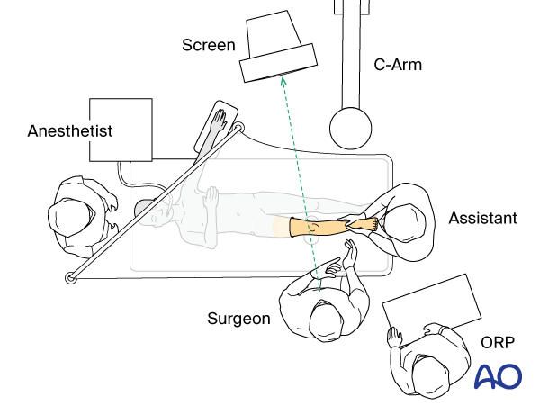
The optimal position of the surgeon is on the injured side of the patient.
The position of the image intensifier screen should allow a direct view for the surgeon.
