Minimally invasive lateral approach to the tibia
1. Principles
Periosteum makes an important contribution to fracture healing in the immature skeleton.
A minimally invasive approach is, therefore, particularly useful in pediatric patients as it causes less damage to the periosteum.
The lateral approach is used for minimally invasive osteosynthesis if the medial soft tissues are injured.
Distally, the shape of the tibia and the overlying soft tissues make it difficult to contour a plate and place screws.
A skin incision sufficient to expose and retract the deep peroneal nerve and anterior tibial vessels is advisable over the distal third of the tibia.
This avoids entrapment of the neurovascular structures by a submuscular plate or injury by percutaneous screws.
2. Anatomy
The triangular shape of the tibia
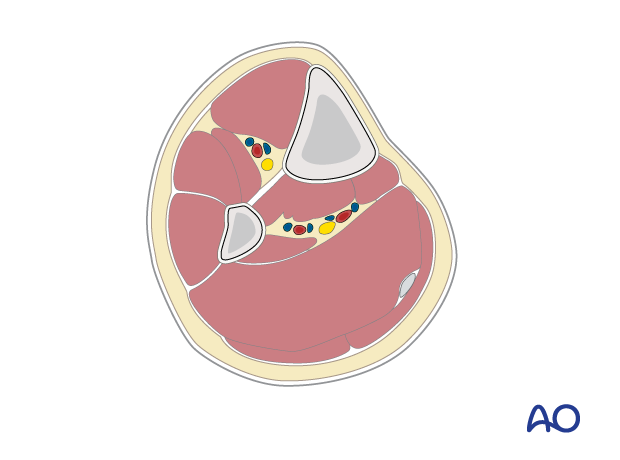
The lateral and posterior surfaces of the tibia are covered by muscle. The anteromedial surface has only a thin layer of subcutaneous tissue and skin. This surface provides less blood supply to the underlying bone.
Compartments
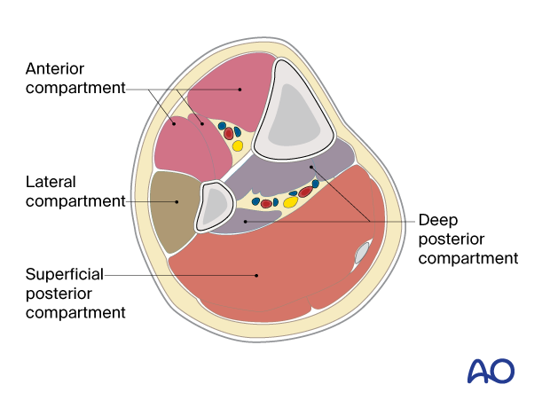
The lower leg has four compartments.
The anterior compartment contains:
- Tibialis anterior
- Extensor hallucis longus
- Extensor digitorum longus
- Anterior tibial artery
- Deep peroneal nerve
The lateral compartment contains:
- Peroneus longus
- Peroneus brevis
- Superficial peroneal nerve
The deep posterior compartment contains:
- Tibialis posterior
- Flexor hallucis longus
- Flexor digitorum longus
- Peroneal artery
- Posterior tibial artery
- Tibial nerve
The superficial posterior compartment contains:
- Gastrocnemius
- Soleus
- Sural nerve
Proximal and distal tibial physes
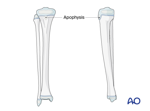
The physis at each end of the tibia must be protected during surgical approaches and implant application.
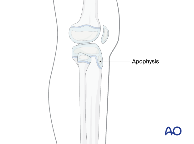
The proximal tibial physis extends anteriorly and distally as an apophysis, which forms the tibial tuberosity.
This may be damaged as a consequence of injury and surgery, leading to tibial deformity.
3. Skin incisions
Identify the level of the physis with image intensification and mark this on the skin before making an incision.
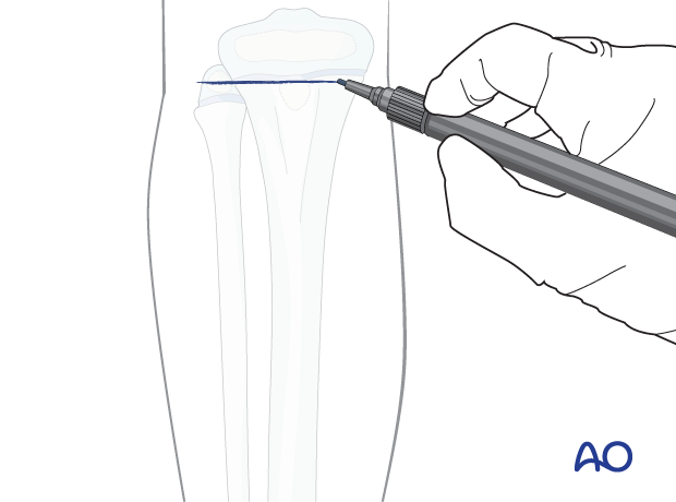
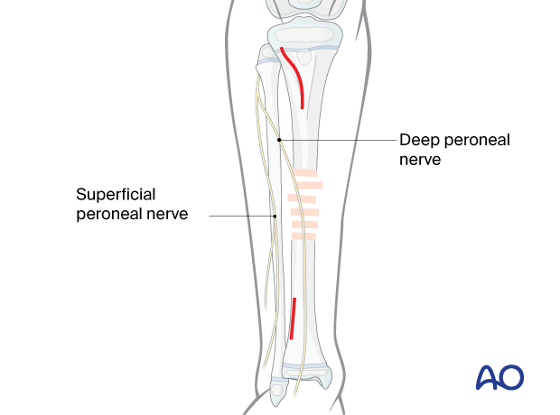
The proximal incision, approximately 5 cm long, begins distal to the tibial tubercle and lateral to the tibial crest. The proximal portion should curve slightly posteriorly.
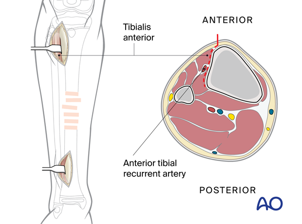
The distal incision is made to prevent neurovascular injury from percutaneous screws. The anterior tibial vessels and deep peroneal nerve cross the anterolateral surface of the tibia approximately at the junction of its third and fourth quarter.

4. Deep dissection
Incise the fascia a few millimeters lateral to the tibial crest.
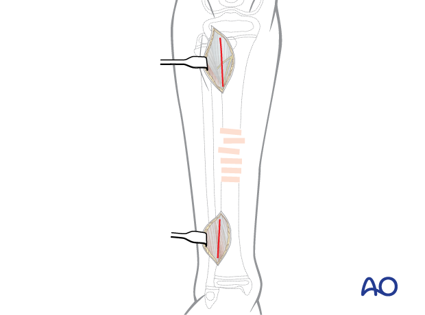
At the distal incision, dissect the tibialis anterior tendon from the bone, preserving the paratenon.
Retract the tendon together with the deep peroneal nerve and the anterior tibial vessels to the lateral side.
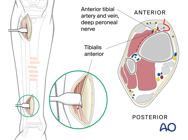
At the proximal incision, dissect the tibialis anterior muscle from the bone without stripping the periosteum or damaging the apophysis.
Retract the tibialis anterior muscle laterally.
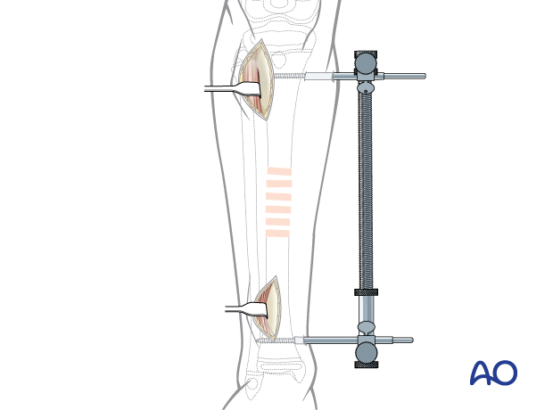
A large distractor helps to reduce and stabilize the fracture.
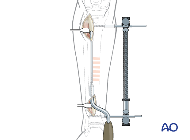
Use a soft-tissue elevator (as shown), or an appropriate plate to prepare a tunnel between the anterior tibial muscles and the periosteum.
Protect the anterior tibial vessels and deep peroneal nerve distally. Depending on how distal the plate is placed, they should be gently retracted laterally or lifted anteromedially so that the tunnel can be placed under the neurovascular bundle, with direct observation.
5. Wound closure
Close the skin and subcutaneous tissues only.


