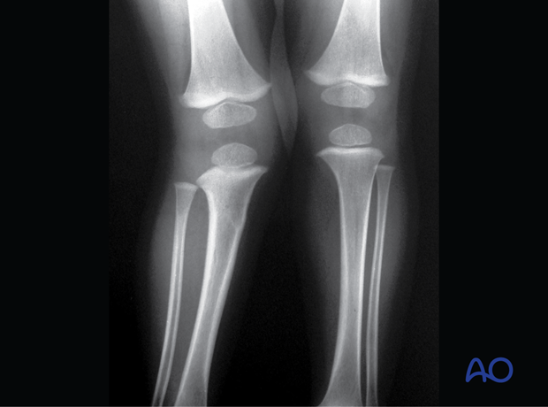
Complete metaphyseal and greenstick fracture
Introduction
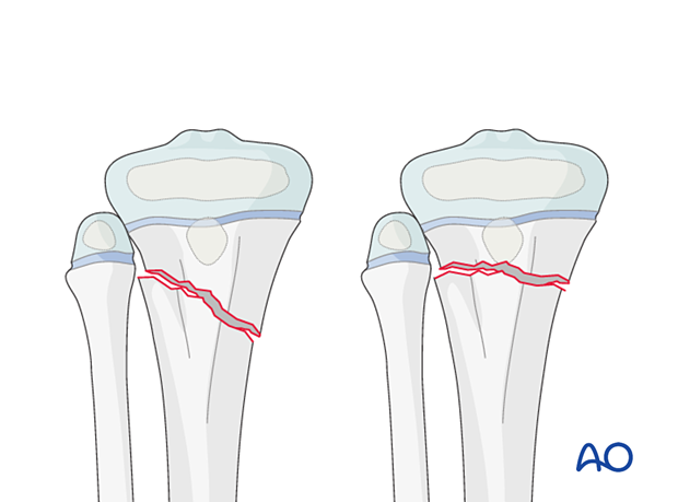
Simple and multifragmentary complete metaphyseal fractures of the proximal tibia are classified as 41t-M/3.1 and 41t-M/3.2.
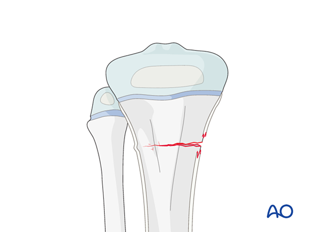
Greenstick fractures involve compression on one cortex and tension/distraction on the opposite cortex.
An associated fibular fracture may have a different configuration, involve the physis, and is classified separately.
Complete fracture
Isolated tibial fractures are often undisplaced.
A transverse fracture is often more stable than an oblique pattern.
Tibial fractures with a fibular fracture at the same level are often unstable.
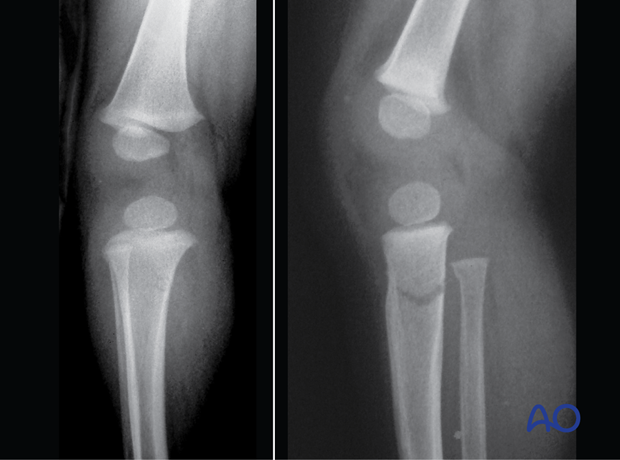
Multifragmentary fractures often result from high-energy trauma and are usually displaced and unstable.
Greenstick fracture
Greenstick fractures are a special type of fracture with compression on one cortex and tension/distraction on the opposite cortex. They are angulated but stable.
Further characteristics
Always consider nonaccidental/unexplained injury in nonambulant children with a tibial fracture.
Soft-tissue injuries are common multifragmentary fractures and should be anticipated. Compartment syndrome and vascular injuries may also complicate these fractures and frequent clinical assessment is mandatory in the early period following the injury.
X-ray
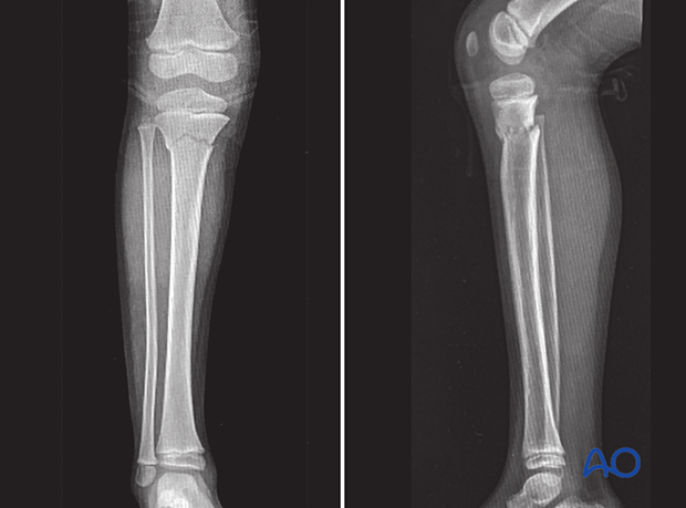
AP and lateral x-rays of a simple metaphyseal fracture of the proximal tibia in a 2-year-old patient
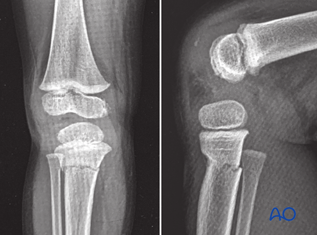
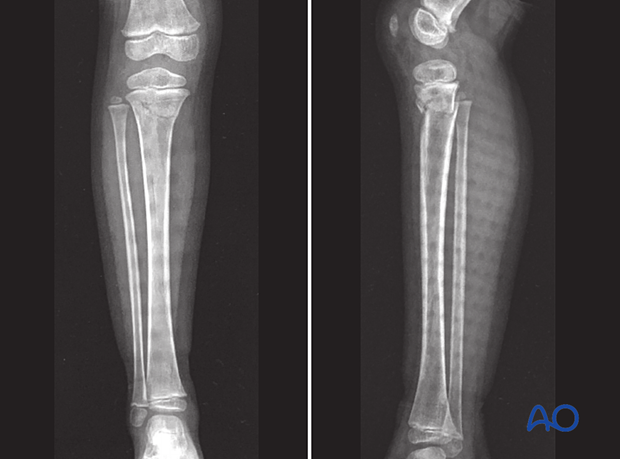
AP and lateral x-rays of another simple metaphyseal fracture of the proximal tibia in a 5-year-old patient
AP and lateral x-rays of a multifragmentary metaphyseal fracture of the proximal tibia in a 4-year-old patient
Cozenʼs phenomenon
Incomplete proximal tibial metaphyseal fractures in young patients can be associated with progressive apex medial (valgus) angulation (Cozen’s phenomenon). When present, this deformity develops in the months following injury and the majority will improve within a few years.
The parents/carers should be alerted to this possibility at the first presentation.
These initial x-rays show an incomplete metaphyseal fracture of the proximal tibia in a 3-year-old child.
In these x-rays 3 weeks postinjury, callus formation can be seen.
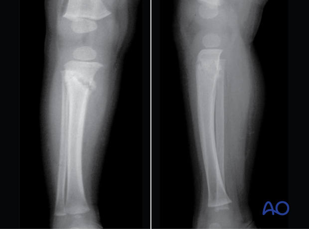
In this x-ray 18 months postinjury, a valgus (Cozen’s) deformity is visible.
