Radiological evaluation
1. General considerations
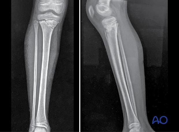
X-rays in two planes are usually sufficient for diagnosis and planning treatment of proximal tibial fractures.
CT/MRI scans may provide useful information in selected cases.
2. Plain x-rays
X-rays in the AP and lateral planes including the knee are required to:
- Identify the location and extent of injury
- Classify the injury pattern
- Evaluate the physes
- Assess the degree of displacement
- Assess stability
- Identify additional injuries
- Identify preexisting contributory pathology
Oblique views may be necessary to evaluate articular and epiphyseal injuries.
3. CT imaging
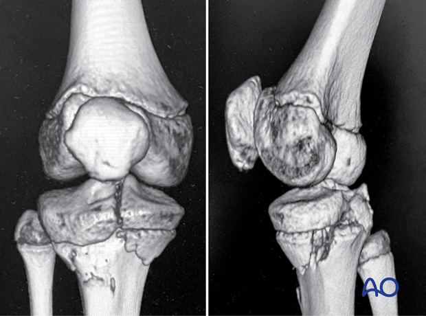
CT imaging is indicated when further information about the fracture morphology is needed for diagnosis and preoperative planning.
In children, smaller (2 mm) cuts should be used to accurately define the fracture pattern.
4. MRI imaging
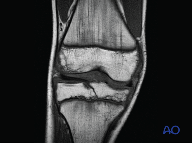
MRI is useful for diagnosing ligament injuries, flake fractures, and for follow-up of physeal injuries to assess the size and extent of bony bridge formation.
This MRI scan (coronal view) demonstrates an intraarticular Salter-Harris type-III fracture of the proximal tibia.
5. Salter-Harris I fractures
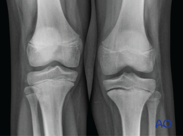
Undisplaced Salter-Harris type-I fractures are difficult to identify radiologically, and the presence of local tenderness and swelling are important signs. MRI may be necessary to confirm the presence of this injury.
6. Nonaccidental/unexplained injuries
All lower limb fractures in nonwalking children should raise concern about the mechanism of injury and require further investigation.
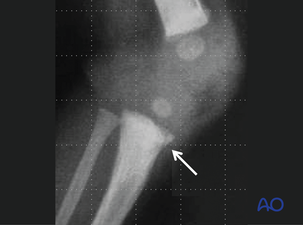
ʻBucket-handleʼ and ʻcornerʼ fractures are fractures with a small metaphyseal fragment in a very young child. This may be caused by traction and rotation and is therefore suggestive of a nonaccidental injury.
