Anatomy of the elbow
1. Introduction
The anatomy of the elbow is complex due to the presence of three major neurovascular bundles and the articular geometry, which is necessary for forearm pronation and supination and elbow flexion and extension.
2. Posterior interosseous nerve
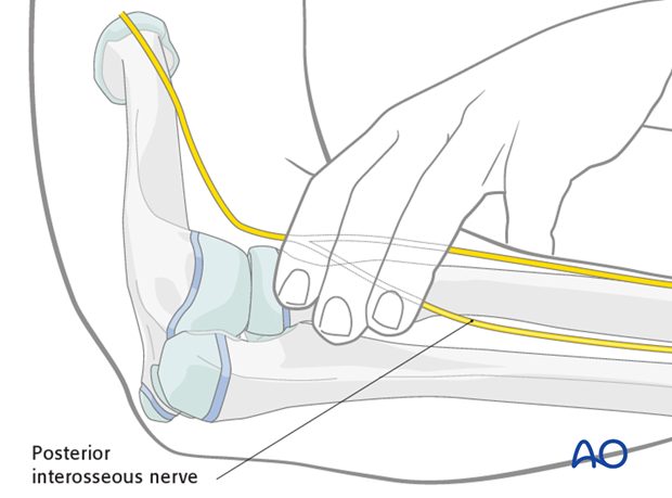
The posterior interosseous nerve (PIN, also known as the deep branch of the radial nerve) is at particular risk during approaches to the proximal radius.
The nerve crosses the proximal radius approximately three patient finger breadths distal to the flare of the radial neck.
Mark this position using three fingers of the child as a reference.
Minimize the risk to the nerve by pronating the arm moving the PIN anteriorly and placing K-wires from a lateral or a posterior direction.
Never place percutaneous K-wires from an anterior direction into the proximal radius.
3. Ulnar nerve
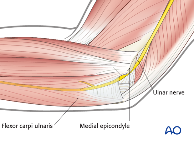
The ulnar nerve lies in the trochlear groove posterior to the medial epicondyle. It passes between the origins of flexor carpi ulnaris and into this muscle at the level of the sigmoid notch of the olecranon.
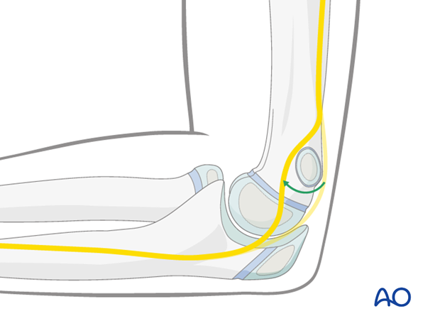
In about 20% of children the ulnar nerve subluxes with flexion onto or anterior to the medial epicondyle. This physical finding can be confirmed on the uninjured limb.
4. Elbow function
The elbow can be thought of as three linked articulations:
- Ulnohumeral
- Radiocapitellar
- Proximal radioulnar
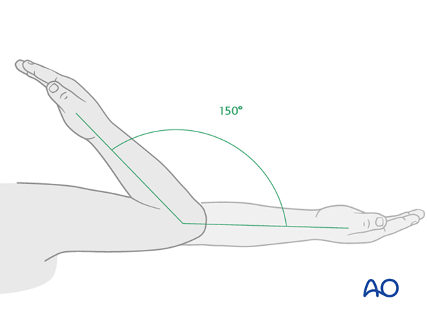
The combined motion of the ulnohumeral and radiocapitellar joints allows 150° of flexion/extension.
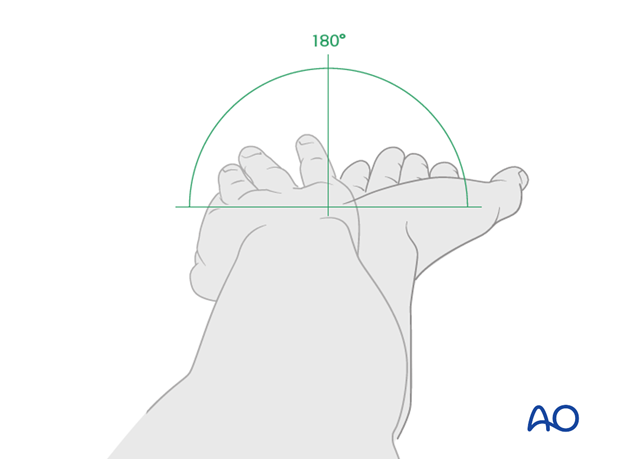
The combined motion of radiocapitellar and proximal radioulnar joints allows almost 180° of pronation/supination.
Restoration of elbow function relies on accurate reduction of radial neck fractures since the radiocapitellar joint participates in both flexion/extension and pronation/supination.
Malalignment of the proximal radius or thickening of the radial neck following fracture healing can reduce pronation and supination. It can also lead to a cam motion with gradual subluxation of the radial head.
5. Developmental anatomy
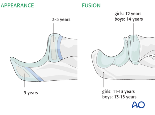
The proximal radius ossification center is the second to appear around the elbow at the age of 3-5 years.
This fuses close to maturity at about 12 years in girls and 14 in boys.
The olecranon ossification center appears at nine years and fuses gradually between 11 and 13 years in girls and between 13 and 15 in boys.
The majority of longitudinal growth in the forearm is from the distal radius and ulna.
Clinical implications: Modeling cannot be relied on to realign malunions of shaft or proximal radius and ulna.
