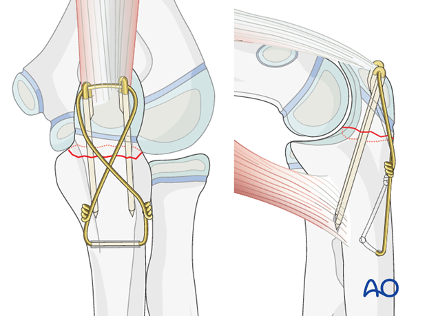
Cerclage compression wire fixation of olecranon fracture
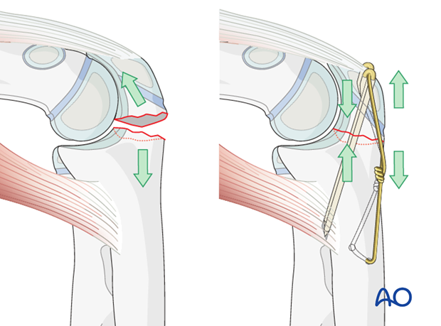
The philosophy behind cerclage compression wiring for the olecranon is that it converts tensile forces on the posterior side of the olecranon into compression forces at the joint line during flexion. The fixation is simple and inexpensive and works well if executed properly.
Choose a wire of sufficient strength to withstand the tensile forces generated in the figure-of-eight loop (1.0 mm). Positioning the K-wires as close to the joint while also penetrating the volar cortex is paramount.
Note: “Cerclage compression wire fixation” was referred to as “Tension band fixation”. We now prefer the term “Cerclage compression wire fixation” because the tension band mechanism cannot be applied consistently to each component of the fracture fixation. An explanation of the limits of the Tension band mechanism/principle can be found here.
1. Instruments and implants
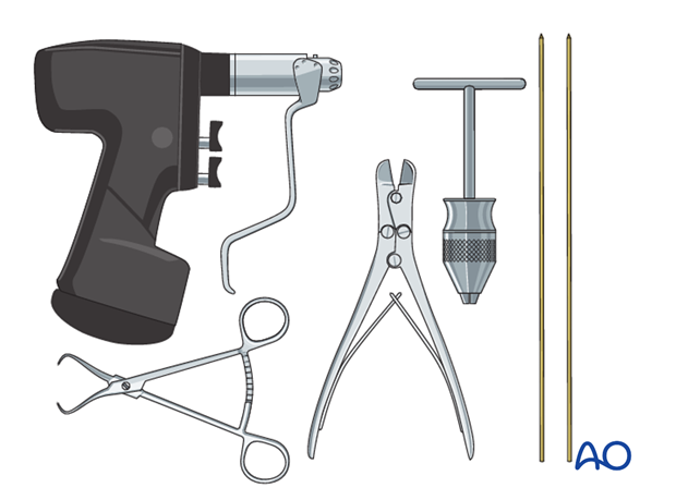
The following equipment is needed:
- K-wires of appropriate sizes
- Wire of appropriate strength
- Drill or a T-handle for manual insertion
- Two pliers for wire twisting
- Wire cutting instruments
- Standard orthopedic instrument set
Alternative: Use a high-strength, braided composite suture or tape instead of figure-of-eight wire.
2. Patient preparation and approach
Patient preparation
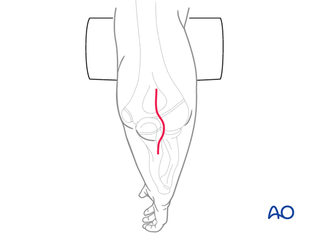
This procedure may be performed with the patient in either a supine position or a lateral decubitus position.
Approach
For this procedure a posterior approach is normally used.
3. Reduction
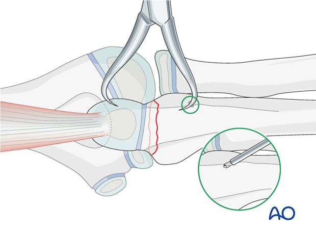
There is almost always a small spike on one of the fragment sides that exactly fits into a gap on the opposite fragment.
Reduce and hold the reduction of the transverse olecranon fracture with a small pointed reduction forceps.
Pearl: Drill a small unicortical hole in the distal fragment to secure the reduction forceps.
4. Prepare wire insertion
Drilling
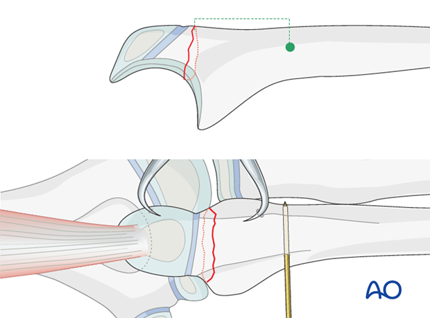
Drill a hole through the ulna approximately 2-4 cm distal to the fracture line and 5 mm from the posterior cortex.
Wire preparation and insertion
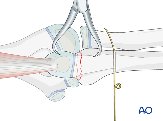
Prepare the wire by making a loop approximately one third along its length.
Insert the shorter segment of the wire through the drilled hole from medial to lateral. As the ulnar nerve is medial it is safer to introduce the wire from this side.
5. Insertion of K-wires
First K-wire
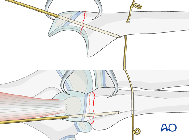
Using the drill guide, introduce the first K-wire medially through the olecranon apophysis.
Aim the drill towards the anterior cortex, passing as close as possible to the joint. Leave enough space on the lateral side for the second K-wire.
Drill the K-wire through both cortices.
Second K-wire
Insert a second parallel K-wire.
Check the position of both K-wires with image intensification.
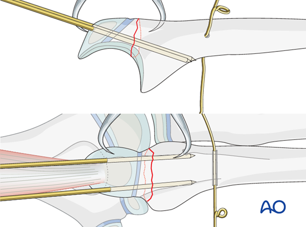
6. Wire fixation
Figure-of-eight configuration
Remove the small pointed reduction forceps.
Pass the long segment of the wire (bearing the loop) in a figure-of-eight configuration beneath the triceps tendon around the protruding ends of the K-wires.
Twist the two wires ends together.
Note: Make sure that the loop and wire twist do not lie too close to the wire hole to avoid unnecessary tension at the wire hole.
Placing the loops more proximally will allow a smaller incision when removing them after healing.
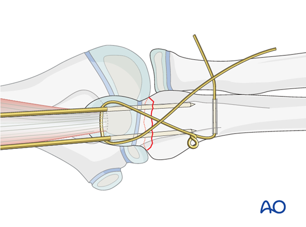
Some surgeons find it easier to use two separate pieces of wire instead of one loop to create the cerclage compression wiring.
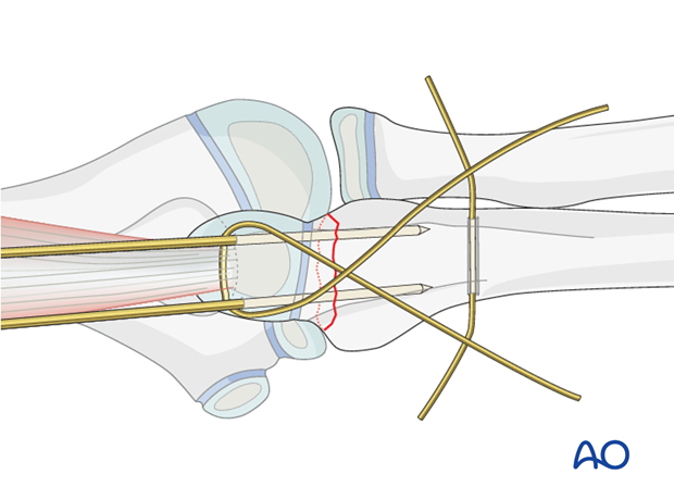
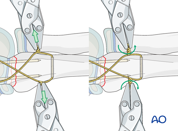
Tightening the wire
Ensure that each end of the wire spirals equally - the twist should not comprise one spiral around a straight wire.
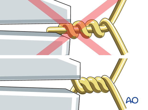
Cut the wire ends short.
The slack is then taken up by further twisting. Repeat this until the desired tension is achieved. Both loops must be tightened at the same time and in the same direction, to achieve equal tension on both arms of the wire.
By tightening the twist and the loop with two pliers simultaneously, the two fragments are drawn together such that the fracture is placed under compression.
Note: Avoid excessive tensioning.
Prevention of later soft-tissue irritation
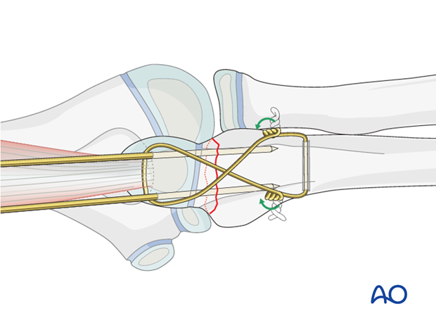
Trim the twisted wire and turn both ends towards the ulna/olecranon to minimize soft-tissue irritation.
Sinking the K-wires
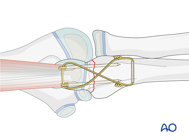
Bend the proximal end of the K-wires 180° with pliers, bending irons or forceps and cut the K-wires.
Sink the curved ends into the olecranon or triceps tendon to prevent backing out and skin irritation.
7. Final assessment
Confirm fracture stability and range of motion, including supination-pronation.
Exclude K-wire penetration into the humeroulnar or radioulnar joint.
Final x-rays or image intensifier views should show good reduction and correct hardware position.
