21r-E/2 Radius, epiphysiolysis with metaphyseal wedge, SH II
General considerations
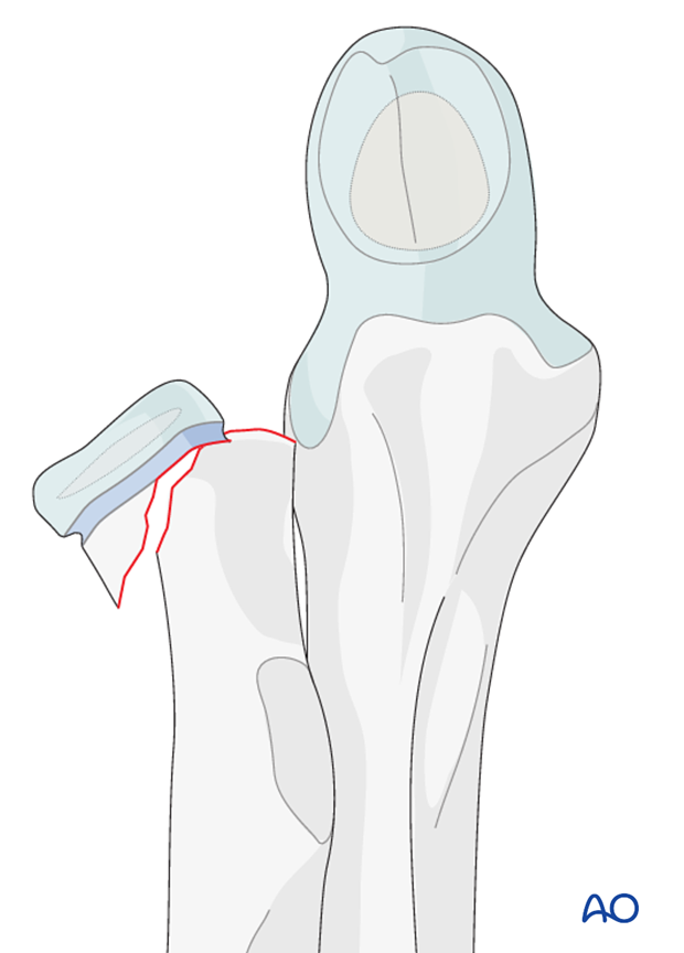
Isolated epiphyseal fractures of the radius with a separation of epi- and metaphysis including a metaphyseal wedge attached to the epiphyseal fragment (Salter-Harris II) are classified as 21r-E/2.
An additional code (I-III) takes into account the axial deviation and level of displacement.
21r-E/2.1 I Epiphysiolysis with metaphyseal wedge, SH II, no angulation and no displacement
Type-I fractures are undisplaced without angulation.

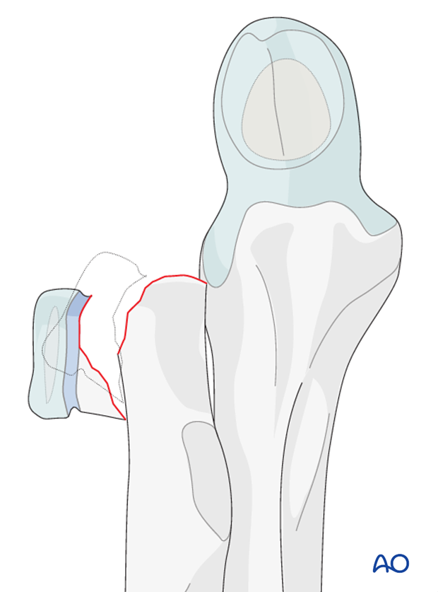
21r-E/2 II Epiphysiolysis with metaphyseal wedge, SH II, angulation with displacement of up to half of the bone diameter
Type-II fractures are fractures with angulation and displacement of up to half of the bone diameter. Displacement is usually lateral. The metaphyseal wedge is usually on the same side as the displacement.
The annular ligament is intact but may be trapped between the fracture fragments.
The head may be impacted in the metaphysis.

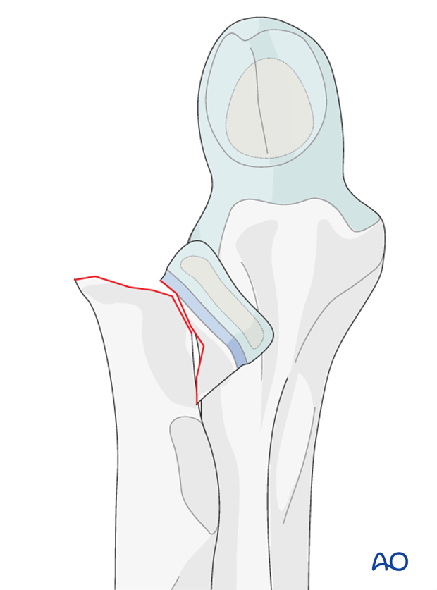
21r-E/2 III Epiphysiolysis with metaphyseal wedge, SH II, angulation with displacement of more than half of the bone diameter
Type-III fractures are angulated fractures with displacement of more than half of the bone diameter. Displacement is usually lateral. The metaphyseal wedge is usually on the same side as the displacement.
The annular ligament is intact but may be trapped between the fracture fragments.
The head may be impacted in the metaphysis.

Medial displacement
This fracture pattern is very rare.
The radial head may be completely displaced medially.
This type of fracture has the highest risk of AVN because of circumferencial disruption of the periosteum.
Take care that the surgical approach does not complete disruption of the periosteum.

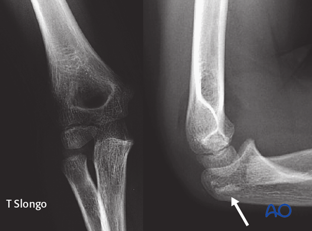
AP and lateral view of a dorsal displacement of the radial head (arrow)


