Vascular anatomy
1. Vascular anatomy
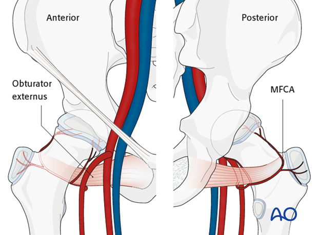
The deep branch of the medial femoral circumflex artery provides the main relevant blood supply to the femoral head.
The medial femoral circumflex artery originates from the deep femoral artery (profunda femoris), courses between the iliopsoas and pectineus muscles, and runs posteriorly between the femur and the pelvis.
During its course, a small branch supplies the inferior retinaculum (ligament of Weitbrecht).
The main branch of the medial femoral circumflex artery is related to the inferior border of the obturator externus muscle and passes posterior to the femur, towards the intertrochanteric crest.
It then crosses posterior to the obturator externus and anterior to the triceps coxae (obturator internus and the superior and inferior gemelli).
Before crossing the triceps coxae, a small branch passes to the greater trochanter.
The vessel enters the joint capsule between the gemellus superior and the piriformis muscles.
Note: The approach must always be cranial to the piriformis muscle. This anatomical detail is crucial when starting to prepare the capsule.
After perforating the capsule, the vessel passes along the superior retinaculum and splits into 3-4 branches.
Provided the obturator externus muscle remains intact, it will protect the medial femoral circumflex artery.


