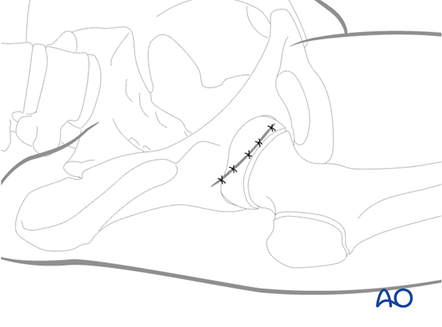
Capsular decompression
1. Preliminary remarks
Introduction
One potential cause of avascular necrosis (AVN) is high intraarticular pressure due to hematoma formation causing intracapsular tamponade. Release of this hemarthrosis may reduce the risk of developing avascular necrosis.
In cases where fixation has been performed without exposure of the fracture, a limited anterior approach is the most direct way to visualize and safely decompress the capsular cavity.
2. Release of joint tamponade
Incision
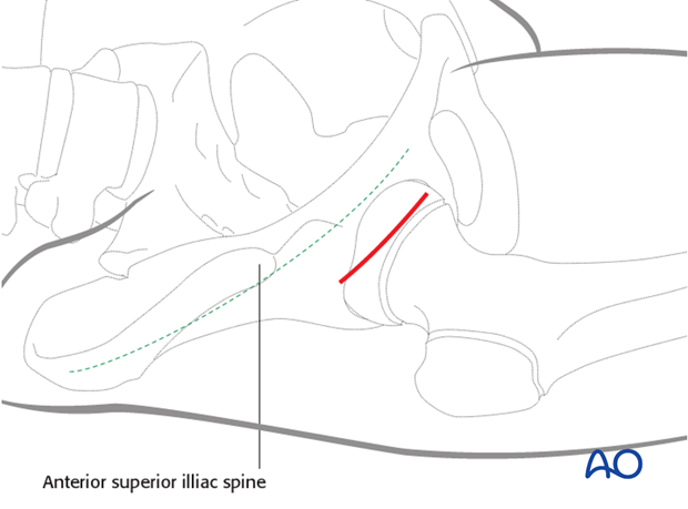
A 2.5 cm incision is made below the anterior superior iliac spine (ASIS) and parallel with the iliac crest. This incision can often be placed in a skin crease for good cosmesis.
Dissection
The incision is deepened to fascia and the interval between the sartorius and the tensor fascia lata is palpated. This interval is easier to identify distally in the wound and becomes confluent as the insertions into the ASIS are approached.
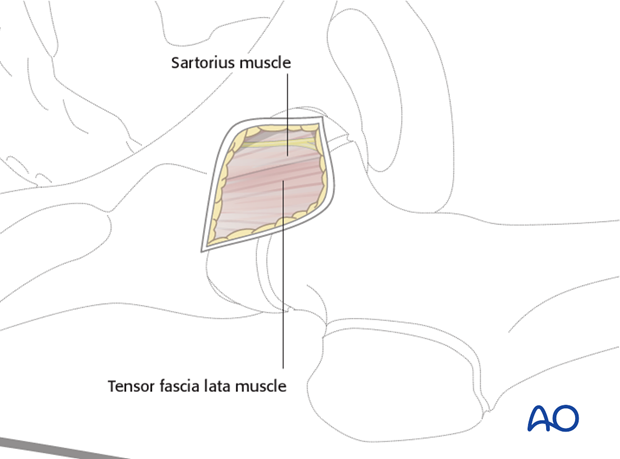
The fascia over the interval is incised with a scalpel and the plane developed with blunt scissors.
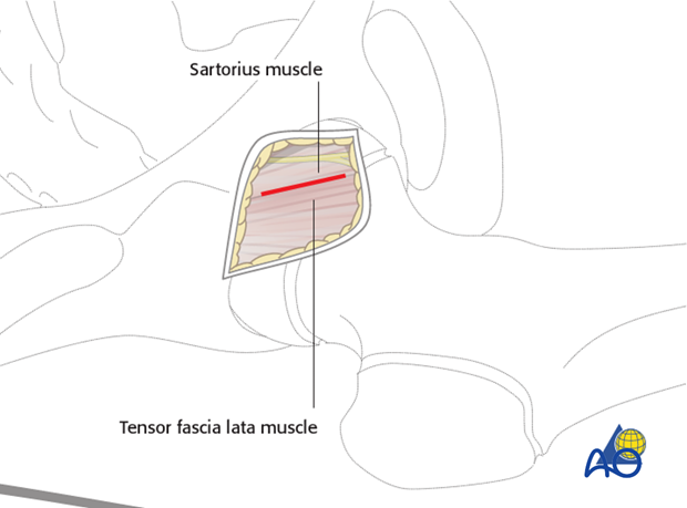
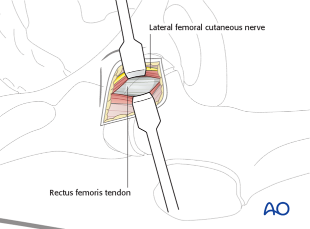
The lateral femoral cutaneous nerve may be identified and retracted medially with the sartorius muscle.
Directly deep to these muscles, the rectus femoris tendon is readily identified. It is round, shiny, and silvery. It can be mobilized and retracted medially exposing the capsule over the femoral neck.
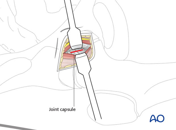
The joint capsule is incised, releasing the tense hemarthrosis.
Closure
The joint capsule is left open. The muscle planes do not need suturing. The subcutaneous tissue and skin can be closed with absorbable sutures.
