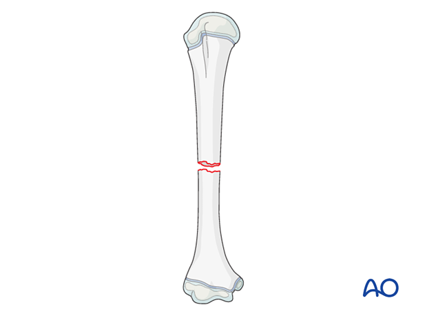
12-D/4.1 Simple complete transverse fracture
Definition
Simple complete transverse or ≤30° oblique fractures of the humeral shaft are classified 12-D/4.1.
Humeral shaft fractures represent 2–5% of childhood fractures.
These fractures are more frequently seen in children 12 years of age and older as a result of falls and sport/recreation injuries, and in children below 3 years of age where the suspicion of a nonaccidental injury should be explored.

Remodeling
The closer the fracture is to the proximal physis the better the remodeling potential.
Translation remodels completely and is independent of location.
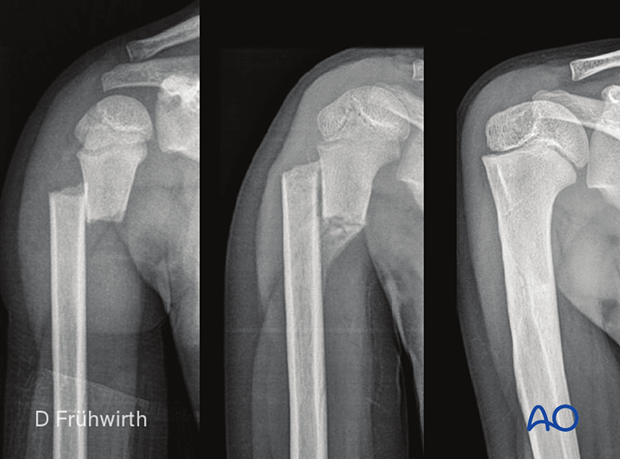
Case of a proximal shaft fracture in a 4-year-old patient, x-rays at injury and 12 months post-injury showing complete remodeling of the translation with nonoperative treatment

Remodeling of varus deformity is more predictable and whilst there is no functional limitation, persistent angulation of >20° can lead to visible deformity.
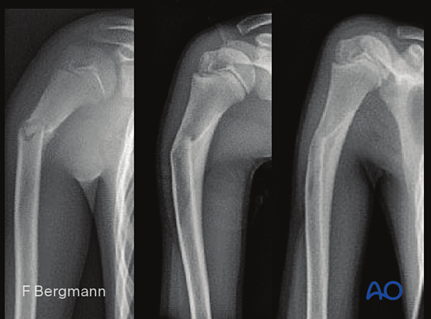
Case of a proximal shaft fracture in a 10-year-old patient, x-rays at injury and 6 months post-injury showing remodeling with nonoperative treatment with subtle residual varus angulation

Radiological evaluation of rotational malalignment is difficult.
X-ray
Most fractures can be identified on plain x-rays.
AP and lateral views including the elbow and shoulder are usually sufficient for diagnosis.
Transthoracic x-rays should be avoided where possible.
A single view including the adjacent joints may be sufficient for surgical decision making.


