The physis
1. The physis
Primary function of growth cartilage
Primary function of growth cartilage is the continuous and controlled elaboration of a solid scaffold (calcified cartilage) in preparation for bone deposition.
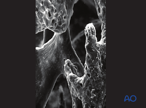
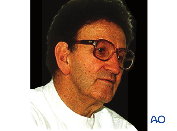
Throughout this discussion of the physis, the images created by the late Professor Robert Schenk of the University of Berne, generously donated to the public domain, via the AO Foundation, have proved an invaluable resource.
Embryological development
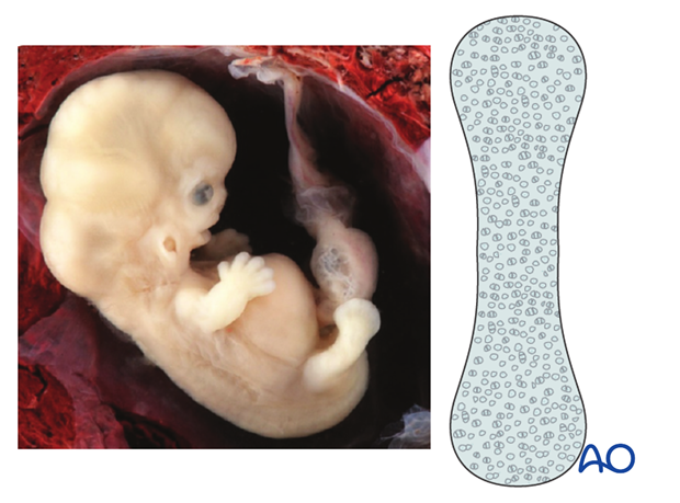
The cartilaginous precursors (anlagen) of the long bones appear early in embryonic life.
The cartilaginous anlage is invaded in its central segment by blood vessels.
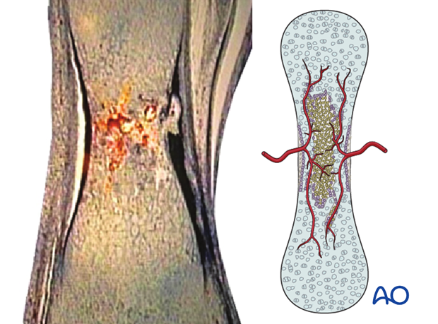
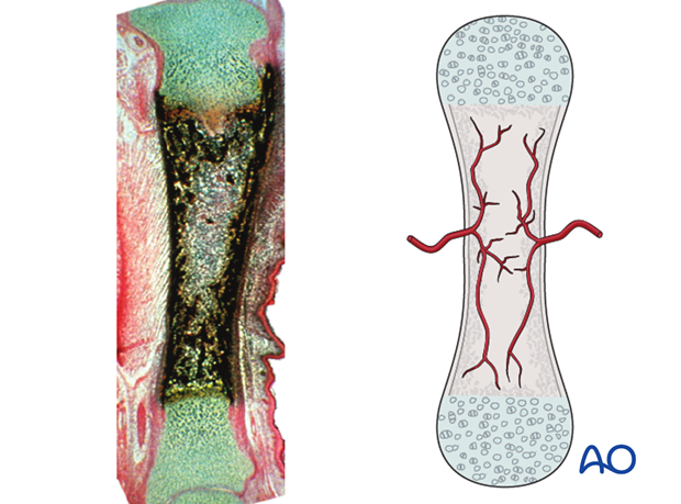
This process results in ossification of the cartilage, producing the early diaphyseal bone.
The histological specimen here is of a young rat’s tibia.
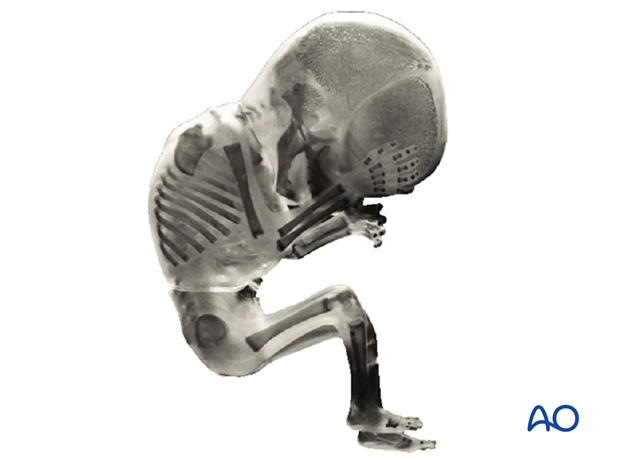
By 13 weeks, in the human fetus, the primary ossification in the diaphyses can be seen clearly.
Postnatal development
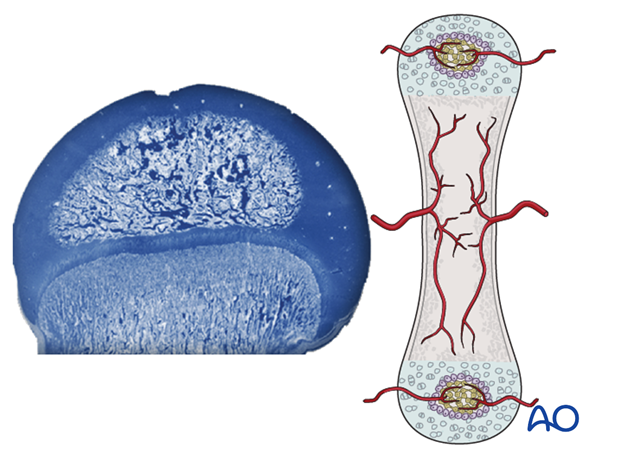
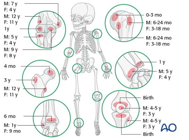
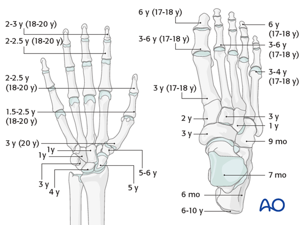
Postnatally, the next skeletal event is the invasion of the cartilaginous end caps (epiphyses) by blood vessels, resulting in the formation of the epiphyseal ossific nuclei (secondary ossification centers).
The times of appearance of the secondary ossification centers of the long bones are shown here.
Similar data for the wrist hand and foot are illustrated here
2. Physeal structure
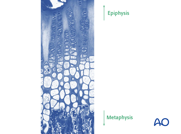
The cartilaginous zones between the bony diaphysis and the ossifying epiphyses differentiate into complex chondral organs – the physes.
Each physis is highly organized into transverse zones
Physeal zones
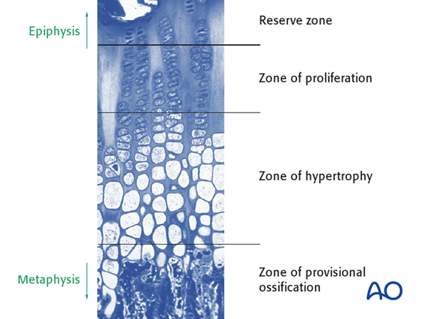
These zones are respectively, from epiphysis to metaphysis:
- The reserve zone
- The zone of proliferation
- The zone of hypertrophy
- The zone of provisional ossification
Each zone contributes different aspects of longitudinal growth.
Vascular anatomy of physis
Trueta, of Oxford, England, first described the vascularity of the physis. He noted that no significant vessel pierced the physeal disc and that there exist no significant anastomoses between the epihyseal and metaphyseal vascular trees within the bone.
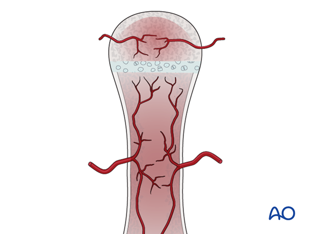
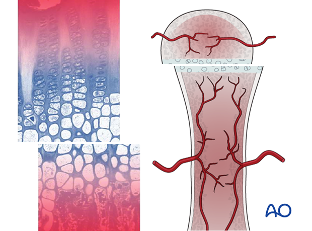
The portion of the physis adjacent to the epiphysis is nourished by diffusion from the epiphyseal circulation and the zone of provisional ossification is supplied by the metaphyseal circulation.
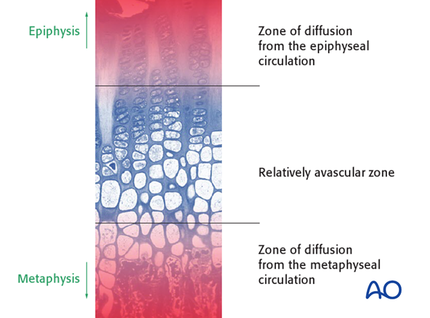
The effect of this is that any shearing disruption of the physis, which usually occurs through the zone of hypertrophy, leaves each of the separated portions of the physis with a functional vascular supply.
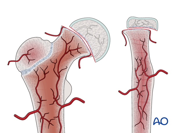
There are some epiphyses that are totally covered by articular cartilage, notably at the proximal femur and the proximal radius, where the vessels feeding the epiphysis are bound down tightly to the perichondrium of the periphery of the physis.
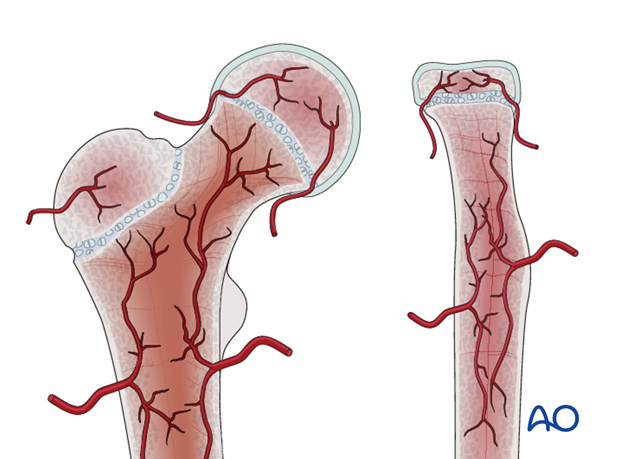
In such sites, a shear injury of the physis is highly likely to devascularize the epiphysis and thereby deprive the reserve and proliferating zones of the physis of nutrition. This commonly results in physeal growth arrest and avascular necrosis of the affected epiphysis.
3. Functional anatomy of growth cartilages
Longitudinal growth
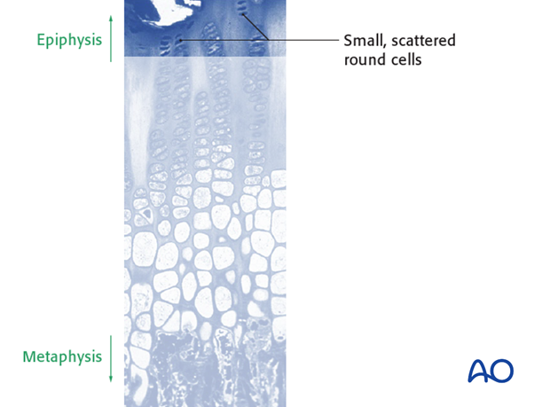
The reserve zone of the physis lies adjacent to the bone of the secondary ossific center: it comprises small, scattered round cells, densely nucleated, and with an abundant endoplasmic reticulum, a clear indication that they are actively synthesizing protein. Their function remains obscure – they do not proliferate at any rate that could contribute to the cell populations of the other zones of the physis.
The proliferating zone of the physis is the zone in which the cells reproduce more rapidly and the more mature ones align themselves in columns, in preparation for hypertrophy.
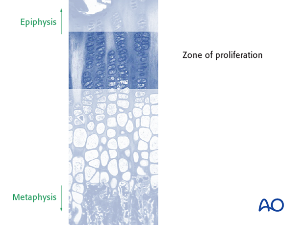
The proliferating zone cells are the fundamental “power-house” of the physis. If they cease to reproduce, for example if deprived of nutrients as a result of lost blood supply to the epiphysis, then longitudinal growth will cease.
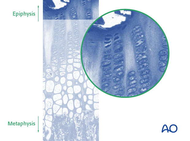
In the hypertrophic zone, the cells are aligned in columns.
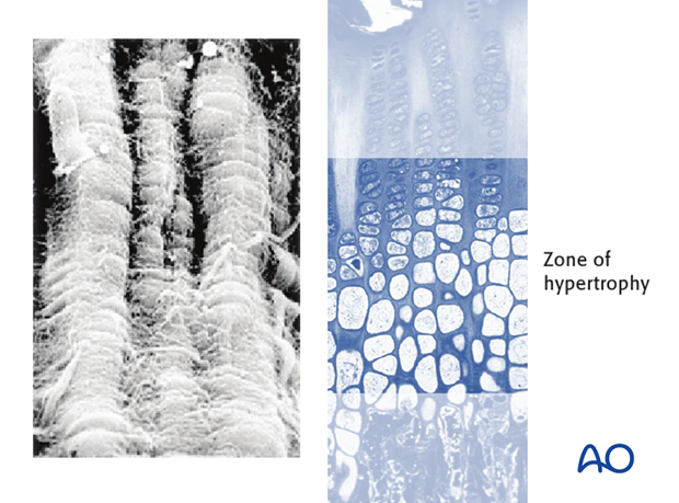
The hypertrophic cells each lie in a lacuna, separated longitudinally by septa and laterally by interterritorial matrix.
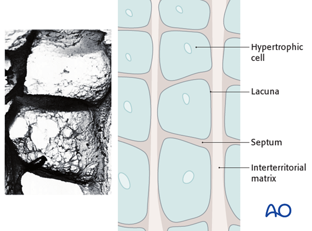
As the cells mature, they enlarge: this enlargement is greater in length than in width and it is this increase in their longitudinal dimensions that is the principle factor resulting in longitudinal bone growth.
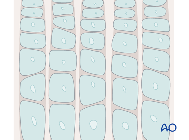
The most mature hypertrophic cells lie adjacent to the zone of provisional ossification.
At this junction the hypertrophic cells undergo programmed cell death – apoptosis.
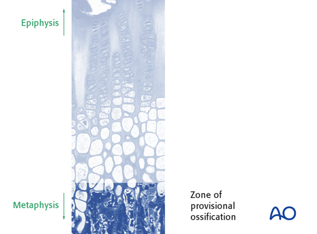
The programmed cell death begins with the ingrowth of capillary buds into the cell lacuna.
Also at this level, the septa and the interterritorial matrix start to calcify.
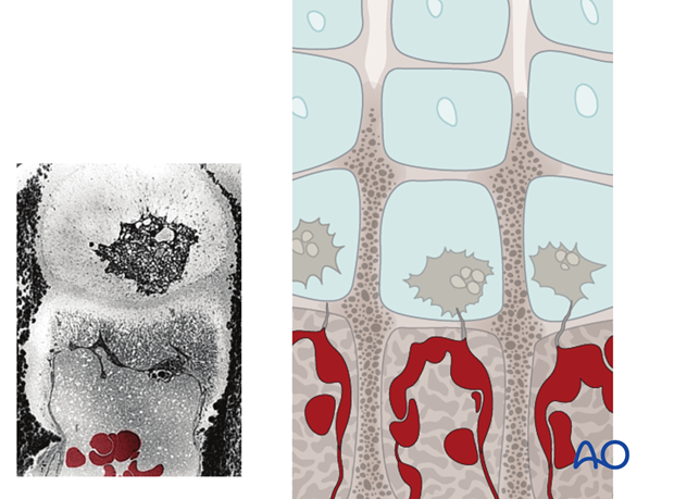
Cells within the walls of the invading capillaries send pseudopodia through the adjacent septum into the lacuna of the most mature hypertrophic cell, which then dies.
This is an active process, triggered by a stimulus that is ill-understood.
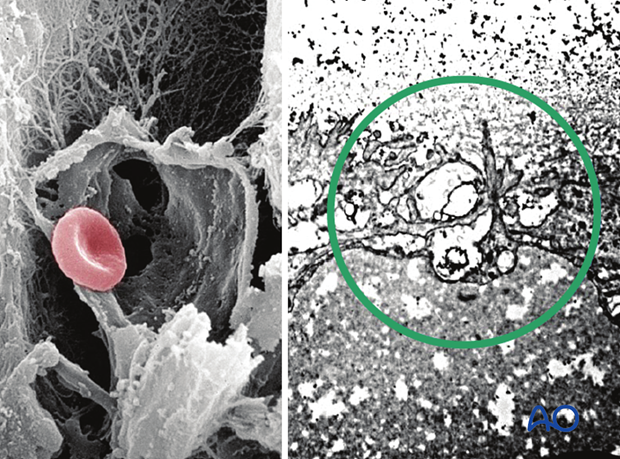
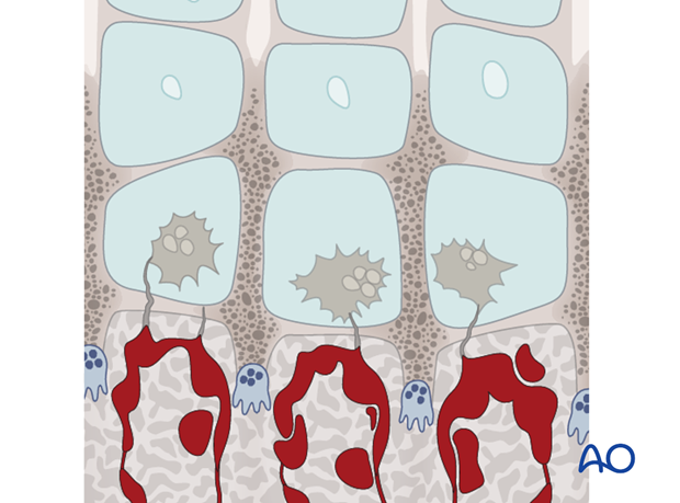
The interterritorial matrix of some of the cylinders occupied by the capillary loops, is broken down by chondroclasts, thereby enlarging the channels.
This specimen, treated with modified von Kossa stain + pyronine, shows the calcified matrix in black. The enlargement of the vascular channels can be seen clearly.
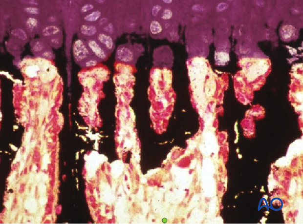
In this preparation, stained with Lovatt’s pentachrome, calcified matrix is green, osteoid tissue is red and primary bone is yellow.
What has happened is that the surfaces of the calcified matrix scaffold have become covered with osteoid tissue that then becomes primary bone, forming the primary trabeculae.
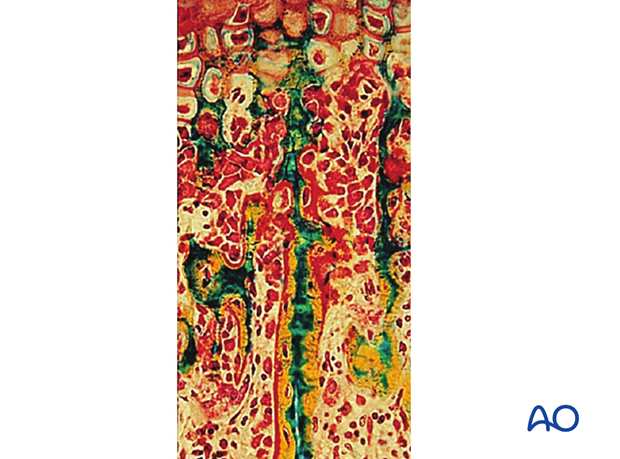
This preparation of a slightly more mature zone of the metaphysis shows the replacement of most of the calcified scaffold (green) by bone (yellow).
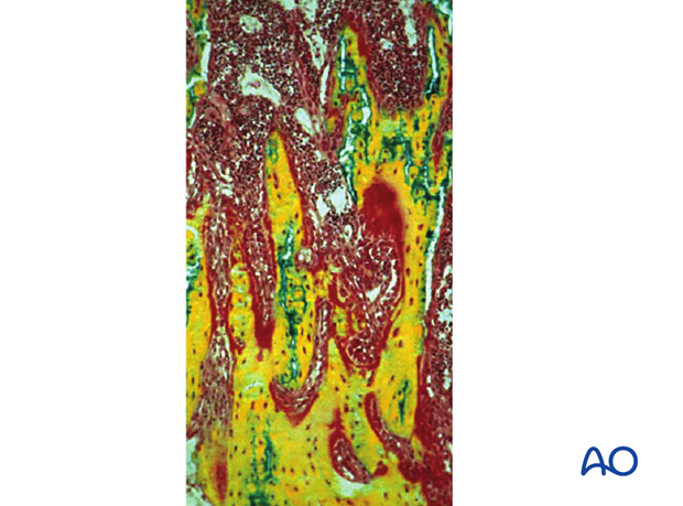
The primary trabeculae then become removed by osteoclasts and replaced by secondary trabeculae, which are orientated according to the stresses of the region.

Diametric growth
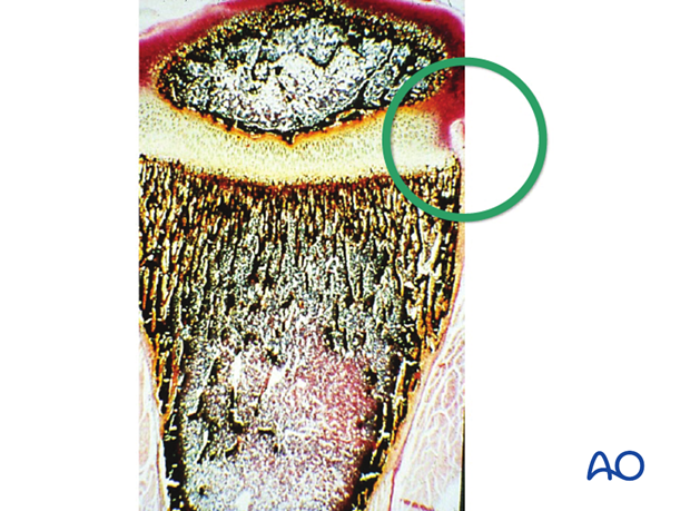
There is a specialized fibrous area surrounding the periphery of the growth plate, comprising the groove of Ranvier and the perichondrial ring of LaCroix. There are cells in this area that are specialized chondrocytes, which increase laterally by appositional growth, thereby resulting in an increase in the diameter of the physis as maturity progresses.
4. Pathoanatomy of injury to growth cartilages
Salter-Harris classification system
The Salter-Harris classification system was first introduced by Robert Salter and Robert Harris in 1963.
See:
- Salter RB, Harris WR. Injuries involving the epiphyseal plate. J Bone Joint Surg Am. 1963 Apr;45(3):587–622.

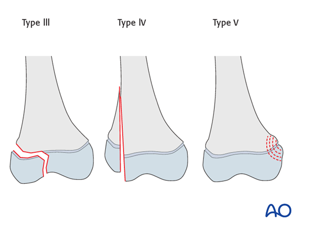
Salter and Harris described 5 patterns of injury to the physis:
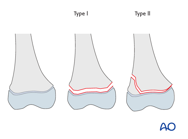
- Type I – a physeal shear without bony injury
- Type II – a partial physeal shear associated with a largely vertical metaphyseal bony fracture
- Type III – a partial physeal shear plus an epiphyseal fracture.
- Type IV – a vertical fracture plane passing through the epiphysis, the physis and the metaphysis.
- Type V – a physeal and metaphyseal crush injury, destroying the related portion of the physis: sometimes evident by a small metaphyseal bulge. Such injuries are frequently diagnosed in retrospect
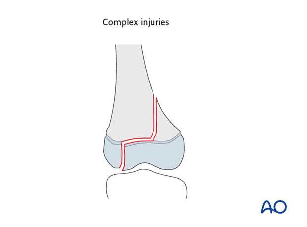
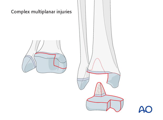
There exist also complex multiplanar injuries of the physis, epiphysis and metaphysis, not categorized by Salter and Harris.
Such complex injuries are exemplified by the triplane group of injuries seen at the distal tibial physis.
There is a validated classification of children’s long bone fractures, developed by the AO Foundation, the result of the work of Dr Theddy Slongo and Dr Laurent Audigé.
