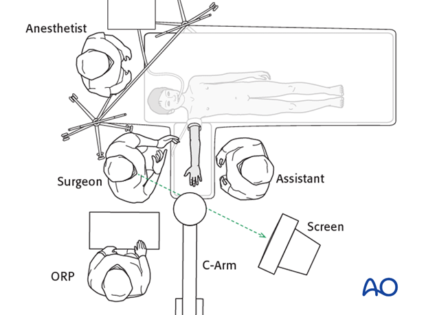
Supine position
1. Preoperative preparation
The following are essential for successful surgery:
- Patient information
- Operating room personnel (ORP) information/instruction
- Surgical planning
2. Patient information
Before treatment discuss the following information with the patient/parents/carers:
- Nature of the injury
- The chosen treatment and why a particular treatment is selected
- Alternative treatments
- General operative risks
- Expected healing time
- Functional recovery
- Implant removal
3. Information for operating room personnel
Operating room personnel (ORP) need to know and confirm:
- Consent form, completed and signed
- Site and side of fracture
- Type of operation planned
- Operative site has been marked by the surgeon
- Condition of soft tissues
- Equipment/implants needed
- Patient positioning
- Duration of operation
- Antibiotic prophylaxis
- Comorbidities, including allergies
4. Surgical planning
The surgeon should ensure that:
- Relevant x-rays and other images are available in the OR.
- Required instruments and implants are accessible and ready.
- Tourniquet is available (a sterile tourniquet makes draping more straightforward).
- Image intensification is available.
- Intraoperative x-ray documentation should be undertaken, with clear AP and lateral views, before applying any plaster cast.
- There is a clear, step-by-step plan of the operation, including backup plans.
5. Anesthesia
- General anesthesia
- Local nerve block
- Combination of nerve block and light general anesthesia
- Prophylactic antibiotics (see local microbiological protocols)
6. Patient position
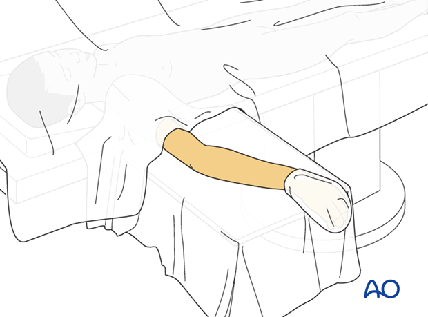
Position the patient supine and place the forearm on an arm table.
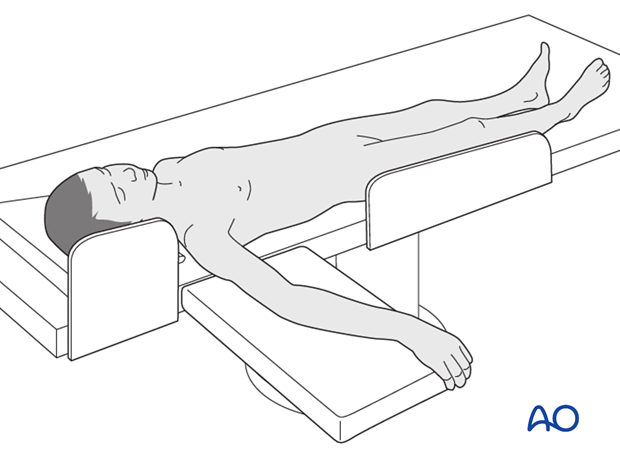
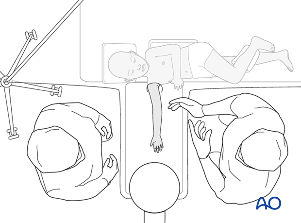
For small children, it may be necessary to rest the shoulder on the arm table to allow unimpeded imaging.
This may require gentle lateral flexion of the neck and trunk with appropriate padding and attention to pressure areas.
By abducting the shoulder, it is possible for the surgeon and the assistant to sit on either side of the hand table.
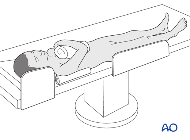
For access to more proximal ulnar shaft fractures, it should be possible to flex the elbow over the child’s chest.
To use the image intensifier the arm must be draped to be able to get AP and lateral views.
7. Skin preparation and draping
With an assistant holding the arm, disinfect the entire arm down to the fingertips.
Drape the hand in a glove or leave it free.
Drape the arm above the elbow.
8. C-arm positioning
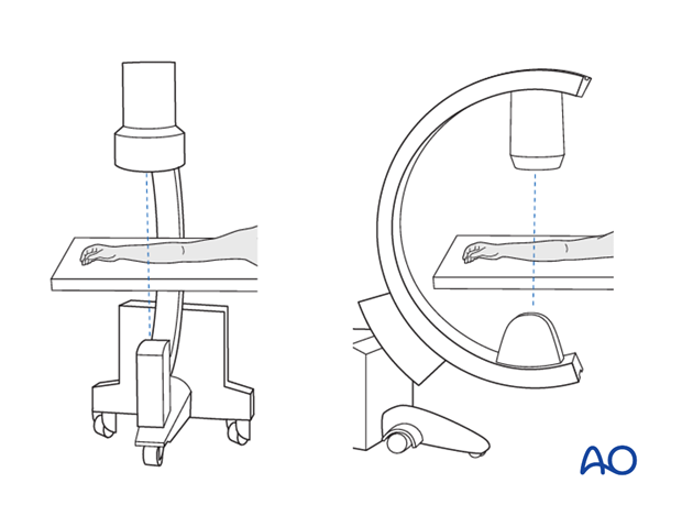
There are two possible positions for the C-arm:
- Parallel to the operating table
- Perpendicular to the operating table
9. OR set-up
The optimal position of the surgeon is defined by anatomical requirements of the fixation and may need to be changed during surgery.
The position of the limb should allow complete imaging of the radius and/or ulna in the frontal and sagittal planes.
The position of the screen should allow a direct view for the surgeon.
The position of operating room personnel should permit ergonomic assistance.
