Patient examination and radiology
1. Visual inspection
The patient is assessed for the following signs:
1. Deformity of the forearm and adjacent joints: Take care to avoid pain during examination.
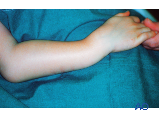
2. Skin discoloration: Indicative of severe fracture displacement and entrapment of muscle/soft tissues
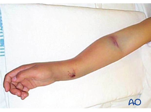
3. Neurovascular examination:
- Peripheral pulses
- Finger movement
- Cutaneous sensation
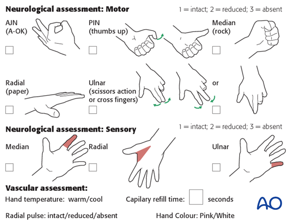
2. Neurological injuries
Neurological injuries in forearm fractures are rare.
Neurological assessment may be difficult and is influenced by the level of pain and the child's age.
3. Vascular injuries
Vascular injuries in forearm fractures are rare.
Peripheral radial pulse is verified by palpation.
Normal pulse does not exclude compartment syndrome.
4. Preliminary reduction
There is no need for preliminary reduction unless the skin is threatened or breached.
Manipulation of a suspected proximal radial fracture may compromise the blood supply to the radial head.
5. Compartment syndrome
Compartment syndrome is an uncommon but devastating complication of forearm fractures.
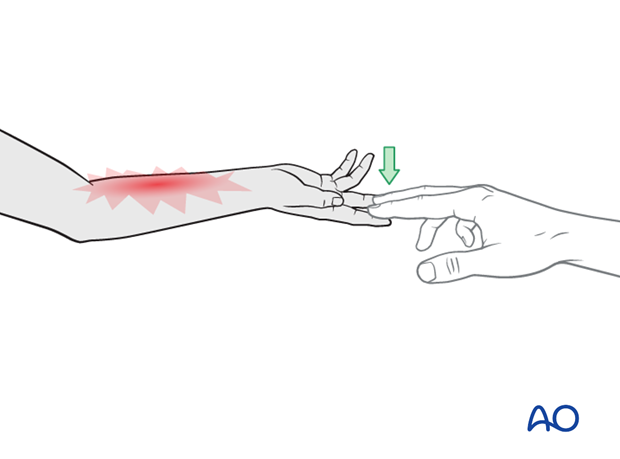
The most straightforward clinical test for evolving muscle ischemia is pain on passive digital extension and/or flexion.
Forearm compartment syndrome and fasciotomy are described in the relevant additional material.
6. X-ray examination of the forearm shaft
Standard AP and lateral views including the adjacent joints are obtained for all forearm shaft fractures.
The arm is often immobilized in a cast making it difficult to obtain standard x-rays.
Suboptimal x-rays are often sufficient for diagnosis and treatment decisions in forearm shaft fractures.
If the fracture is clearly displaced and surgery is indicated, a further view may be postponed until the patient is under general anesthesia.
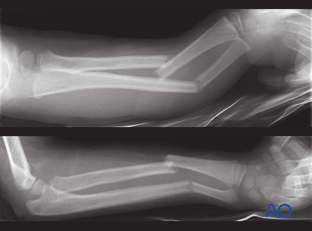
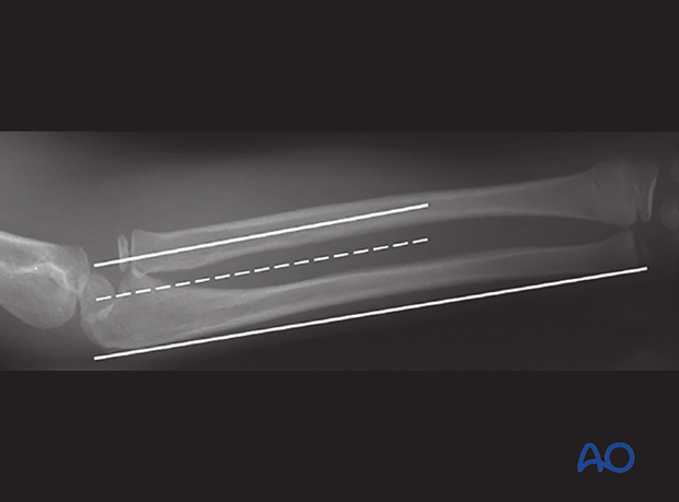
In this example the radiograph does not include both adjacent joints.
Further radiographs are essential to evaluate the injury...
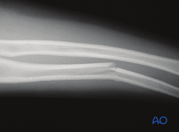
...and in this case identifies dislocation of the radial head.
7. Commonly overlooked fracture patterns
Check for:
- Bowing deformity
- Dislocation of radius (in case of ulnar fracture)
Deviation from a straight line on AP or lateral view may indicate a bowing deformity.
If reasonable doubt exists consider comparison with the uninjured forearm.
Clinical correlation is important. If the arm looks bowed it is.
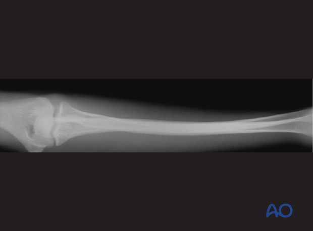
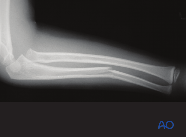
If an ulnar fracture is present, always carefully assess the proximal radio-ulnar joint to identify dislocation of the radial head (Monteggia lesion).
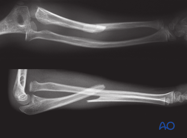
In this case, an ulnar bowing deformity is associated with a dislocation of the radial head.
A straight line drawn along the mid radial diaphysis must intersect with the center of the capitellar ossification center in the AP, Lateral and oblique view
If this association is not present, dislocation of the proximal radio-ulnar joint must be suspected.
