Radius, multifragmentary fracture: open reduction, bridge plating
1. Introduction
General considerations
Plating is the standard technique for treating forearm fractures in adults and is therefore best considered for skeletally mature or nearly mature children.
Children with open physes have thick active periosteum favoring stability and rapid healing with the ESIN method. Where such techniques are unavailable plating may be used in younger children.
If technically possible, ESIN is biologically favored. If plating is used, soft-tissue and periosteal stripping of the bone should be minimized.
Plating of multifragmentary forearm shaft fractures
Plating of pediatric forearm shaft fractures follows the technique for plate fixation in adults.
For multifragmentary forearm shaft fractures, bridge plating may be applied. This results in relative fracture stability.
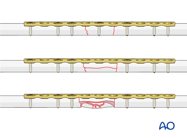
2. Principles
Instruments and implants
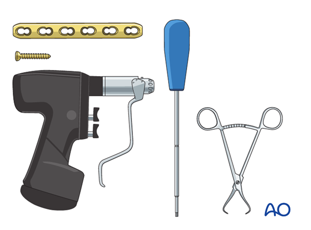
A small fragment set consists of:
- 2.7 or 3.5 mm plates and screws
- Power driver
- 2.7 or 3.5 mm insertion set
Choice of implant
The requirements for internal fixation of complex multifragmentary forearm fractures include:
- The plate should span any comminuted zone and provide at least a three-hole segment over each main fragment, with at least two bicortical screws in each main fragment.
- Well-spaced screws to distribute the stresses on the bone-implant composite for each main fragment
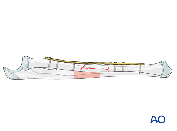
Plate position
Depending on the surgical approach, the plate will be applied to either the anterior or posterior surface of the radius.
In the following example, a plate applied to the anterior surface is illustrated.
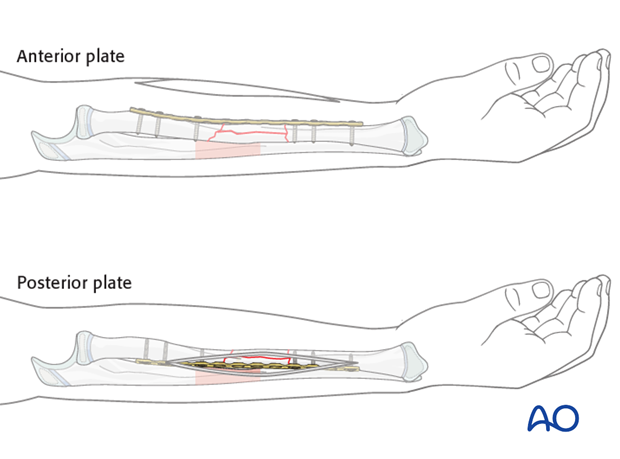
Plate contouring
The plate may need to be contoured to conform to the morphology of the radial shaft.
Choice of approach
For proximal radial shaft fractures, the anterior approach (Henry) is most often used to minimize the risk of damage to the posterior interosseous nerve, which crosses the proximal radius within the supinator.
In mid and distal radial shaft fractures, either the anterior approach (Henry) or posterolateral approach (Thompson) can be used, depending on surgeon’s preference.
3. Reduction
Reduction and fracture stability in children
Children’s periosteum is thick, tough tissue and is often intact on the concave (compression) side of a fracture.
Reduction maneuvers should be gentle to take advantage of stability of an intact periosteum.
Exaggeration of the fracture deformity may be required to loosen the periosteum and allow for gentle reduction.
In pediatric fractures there is often a combination of patterns of bone failure. Residual plastic deformity may prevent anatomical reduction of some of the fracture edges. Provided the alignment of the bone is anatomical and overall reduction is stable it is not necessary to perfectly reduce the entire fracture.
Open reduction
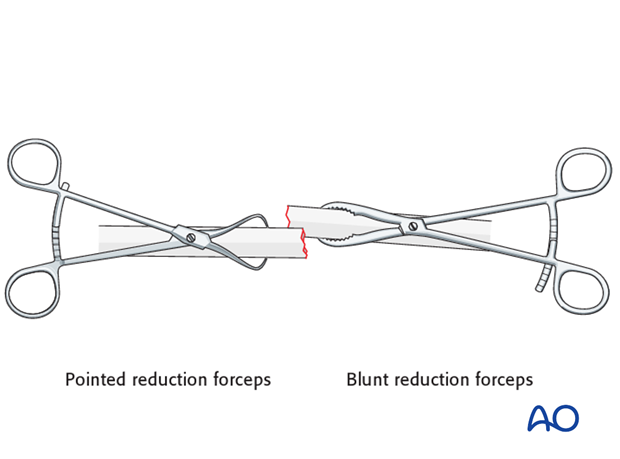
Reduce the fracture without extensive dissection of the fracture zone, using a reduction forceps on each main fragment.
The use of blunt, as opposed to pointed, reduction forceps can be helpful, particularly if greater forces are required.
Fix the plate to one fragment and then reduce the other main fragment onto the plate maintaining length, alignment and rotation.
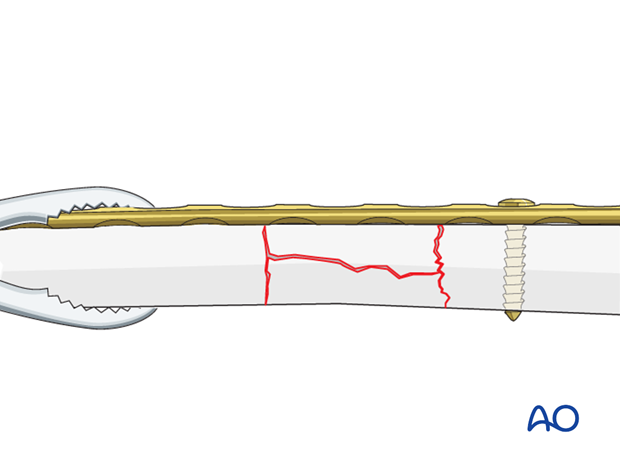
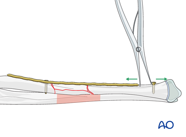
Plate assisted reduction and distraction using a laminar spreader and a screw (push-pull technique)
The plate is fixed to the proximal main fragment using a standard screw though the end hole.
Check that the rotational alignment is correct.
An independent screw is inserted into the distal main fragment a short distance (1-1.5 cm) beyond the distal end of the plate.
Use a laminar spreader between the independent screw and the end of the plate to distract the fracture, restoring length and axial alignment.
4. Plate length and number of screws
Three bicortical screws are suggested in each main fracture fragment due to the high torsional stresses.
Depending on its size, the intermediate fragment(s) may be fixed with bicortical screws or spanned by the plate, remembering that bridge plating is to achieve relative stability.

5. Fixation
Plate positioning
Make sure that the plate is seated on the bone without any soft-tissue interposition apart from periosteum which should not be stripped.
In rare cases, the posterior interosseous nerve is identified. In such cases it is recommended that the position of the nerve in relationship to the plate be noted and documented in the operation report.
Insertion of 1st screw
Align a suitable plate to the bone, keeping in mind the shape of the yet unreduced fracture fragment.
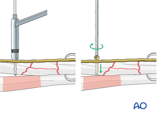
Insert a neutral screw through the plate into one of the main fragments near the fracture line.
Insertion of 2nd screw
Insert a screw through the plate onto the other main fragment.
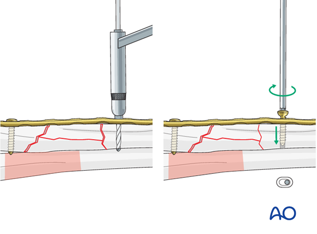
Optional: Fixation of intermediate fragment(s)
Secure the intermediate fragment(s) to the plate with a screw(s) if appropriate.
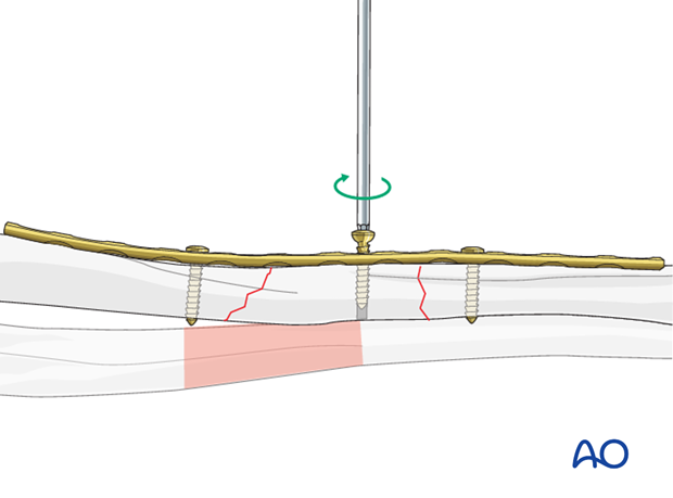
Completed osteosynthesis
Before inserting additional screws check reduction of the fracture and rotation of the forearm.
Insert the remaining screws.


