ESIN entry points (pediatric ulna)
In the ulna use either the proximal lateral or the distal medial entry point.
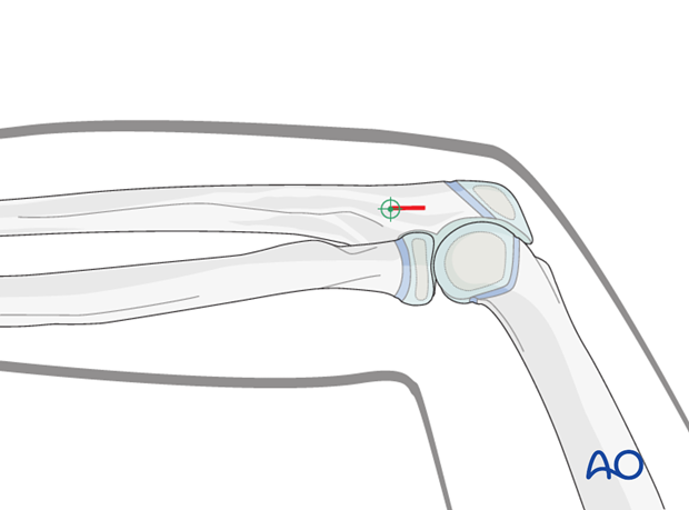
1. Proximal lateral (radial) entry point for the ulna
Determine the correct entry point by palpating 1 cm lateral to the dorsal ulnar rim and 3 cm from the olecranon tip and verify it with an image intensifier.
Perform a stab incision down to the bone.

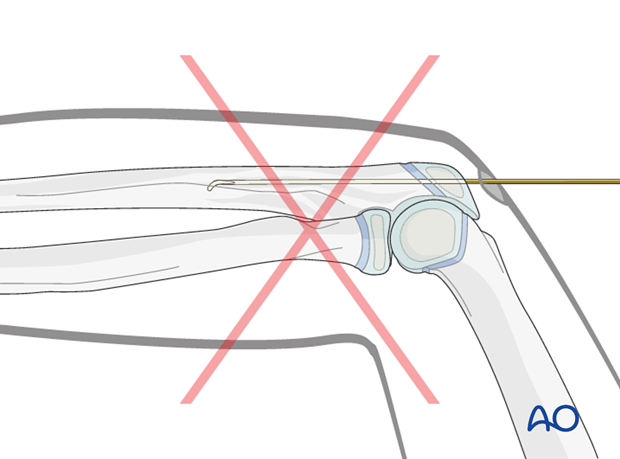
Pitfall: Entry point through the olecranon
Do not introduce the nail through the olecranon.
This does not enhance fixation and causes troublesome soft-tissue irritation and discomfort to the child.

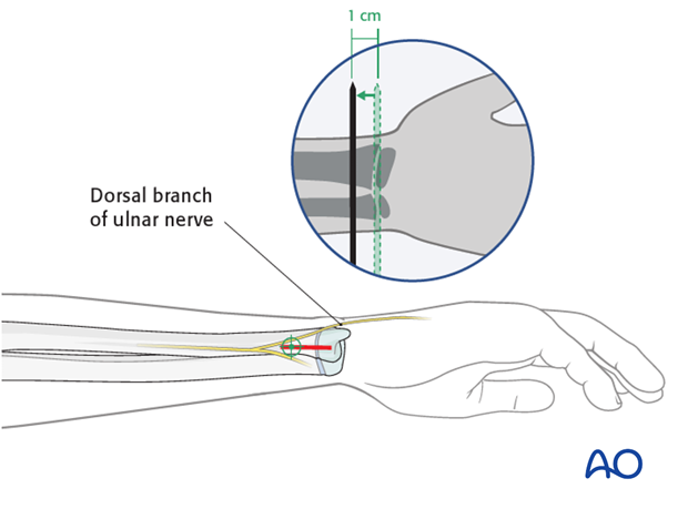
2. Distal medial entry point for the ulna
Use an image intensifier to determine the correct entry point.
This lies on the medial side of the ulna, 1 cm proximal to the growth plate.
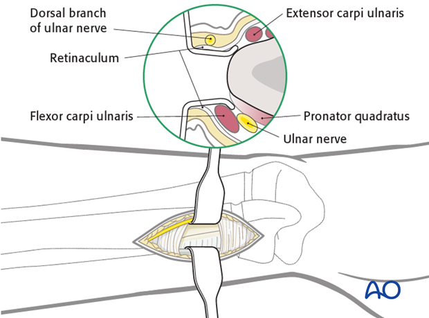
Start the skin incision at the level of the entry point and extend it 2 cm distally.
If an image intensifier is not available, it is safer to use the proximal entry point.

Use small scissors or a surgical clip and small retractors to dissect to the bone under direct vision.
Be careful not to injure the dorsal branch of the ulnar nerve or the basilic vein.

3. Wound closure
Close skin and subcutaneous tissue with fine resorbable sutures (this avoids distress to the child when removing nonabsorbable sutures).


