Approach to the pediatric ulna
1. Introduction
The standard ulnar approach offers good exposure along the whole ulnar shaft. The length of the incision depends on the exposure needed.
In the following, the approach to the middle and distal thirds of the ulnar shaft are illustrated. This approach can be extended both proximally and distally.
2. Skin incision
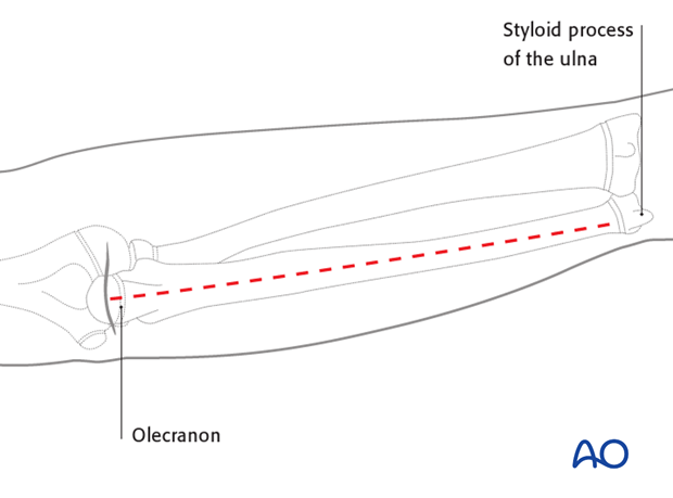
Incise the skin following the subcutaneous border of the ulna, along a line drawn between the tip of the olecranon process and the ulnar styloid process.
Pearl: If the forearm is markedly swollen, it may not be possible to close the skin of the ulnar approach.
In these circumstances, it is better to plan the skin incision over the adjacent extensor muscle compartment, so that an open incision will have a muscular bed rather than exposing the implant.

3. Deep dissection
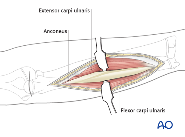
Perform a deep dissection in the interval between the flexor carpi ulnaris and the extensor carpi ulnaris.

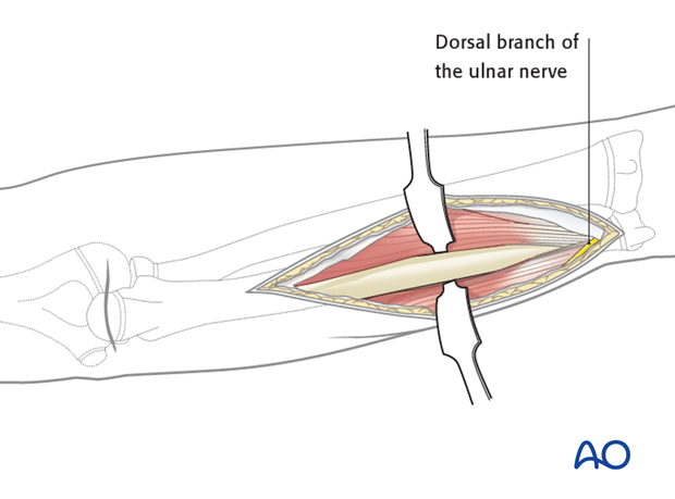
Note: In a very distal extension of the ulnar approach, take care to avoid injury to the dorsal branch of the ulnar nerve, as it runs to the dorsum of the hand.

4. Wound closure
Generally, the muscles do not need reattachment as they fall back to the anatomical position. It may be appropriate to tack the flexor carpi ulnaris to the extensor to cover any implant. The fascia should not be closed after forearm approaches because of the risk of compartment syndrome.
Close skin and subcutaneous tissue with fine resorbable sutures (this avoids distress to the child when removing nonabsorbable sutures).


