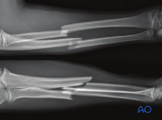
22-D Both bones, combination of radial and ulnar fracture
General considerations
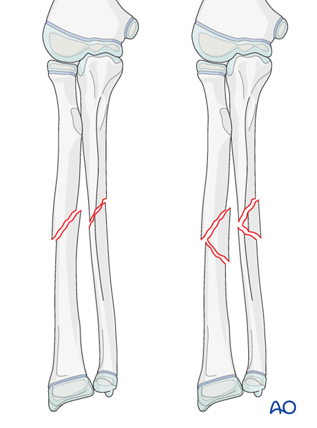
Mid-diaphyseal fractures of each or both bones are illustrated. In practice, many individual fracture patterns occur.
Follow the links in the procedures for reduction and fixation of each individual bone.
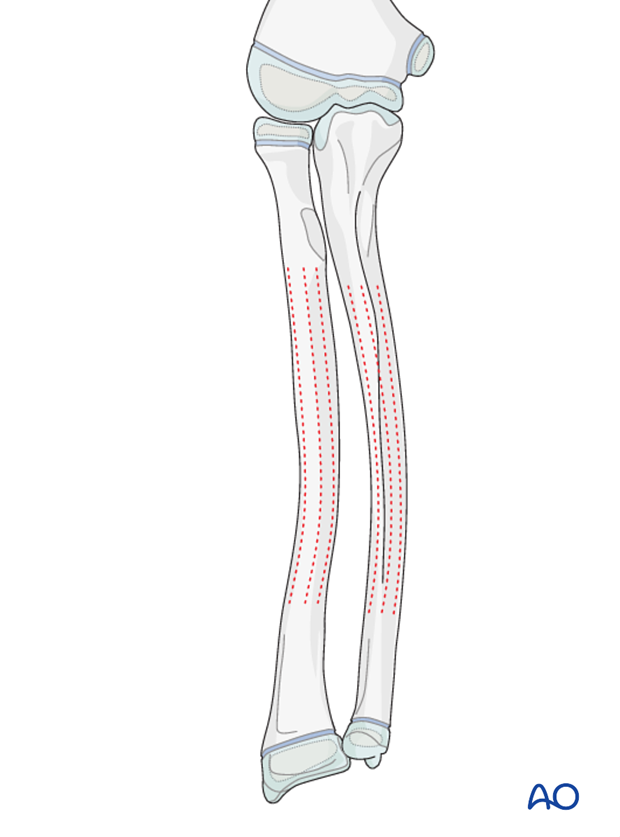
22-D/1.1 Both bones, bowing
Traumatic bowing is a plastic deformity of the radius and ulna. The cortical bone is subject to tensile forces on the convex side and compressive forces on the concave side. If this exceeds the elastic limit, but not the load to failure, deformity will occur without radiological evidence of fracture.
Bowing is more common in young children or in older children with vitamin D deficiency.
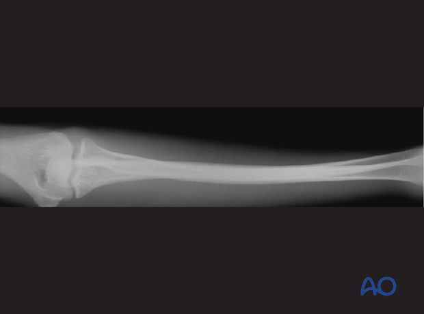
Plastic deformity of a long bone, especially in the forearm, can easily be overlooked. Knowledge of the radiological anatomy and correct axis of each bone is therefore important.
An unreduced plastic deformity of both bones is often associated with loss of pronation and supination.
22-D/2.1 Both bones, greenstick fracture
A greenstick fracture is a combination of a complete fracture on the tension (convex) side and plastic deformity on the compression (concave) side.
The periosteum is disrupted on the convex side and remains intact on the concave side.
Greenstick fractures occur mostly in young children.
The location of this fracture combination is usually at the same level.
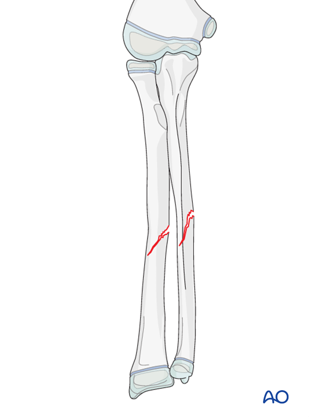
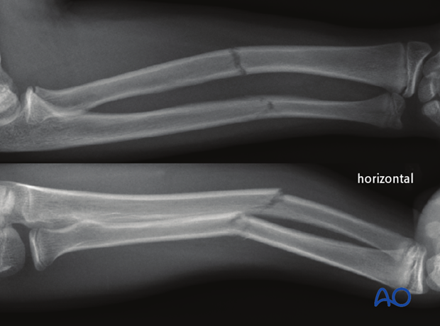
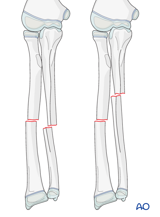
22-D/4.1 Both bones, complete transverse (≤30°) fracture, simple
Simple, complete transverse fractures of both bones may be at the same or different levels.
This has an impact on initial and post-reduction angular stability and influences the choice of treatment.
Instability is common in:
- Completely displaced fragments
- Fractures at the same level
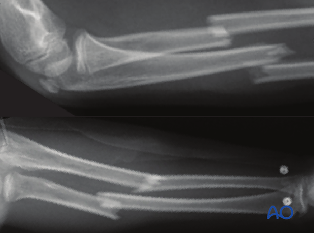
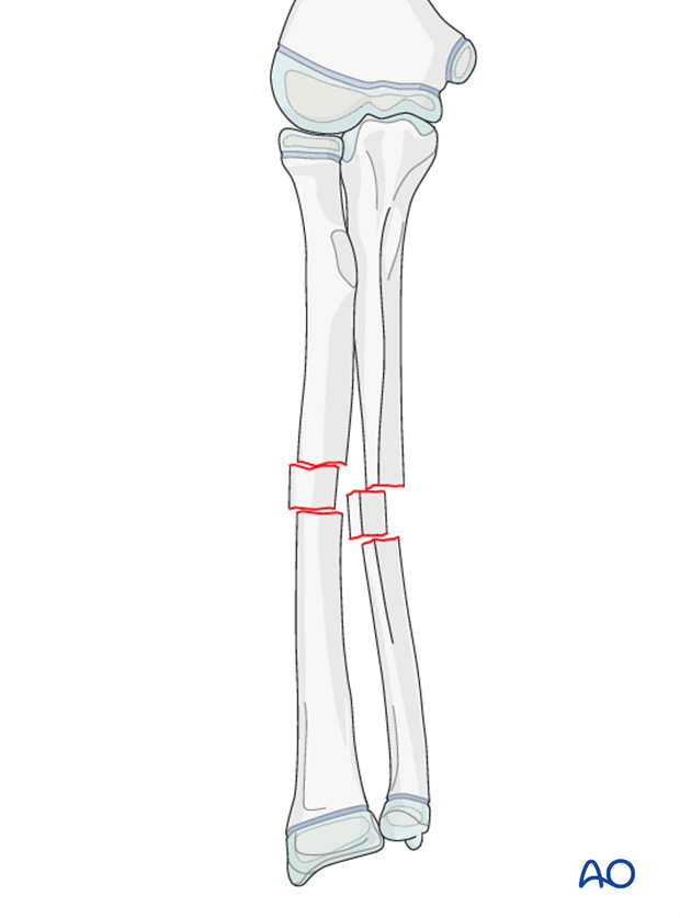
22-D/4.2 Both bones, complete transverse (≤30°) fracture, multifragmentary
Multifragmentary, complete transverse fractures are inherently unstable.
22-D/5.1 + 5.2 Both bones, complete oblique or spiral (>30°) fracture, simple and multifragmentary
Complete oblique or spiral fractures are inherently unstable.
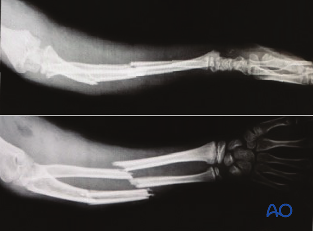
Both bone injuries in combination with radial head dislocation
Both bone fractures can occur in association with radial head dislocation. This is referred to as a Monteggia lesion.
It is therefore mandatory to include both adjacent joints in forearm shaft x-ray images.
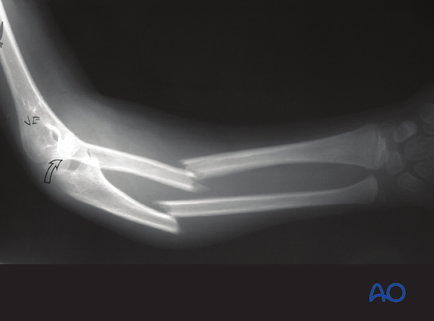
Both bone injuries with combination of fracture types
Greenstick fracture of the radius and bowing of the ulna
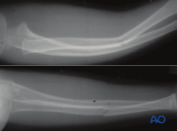
Complete transverse fracture of the radius and bowing of the ulna
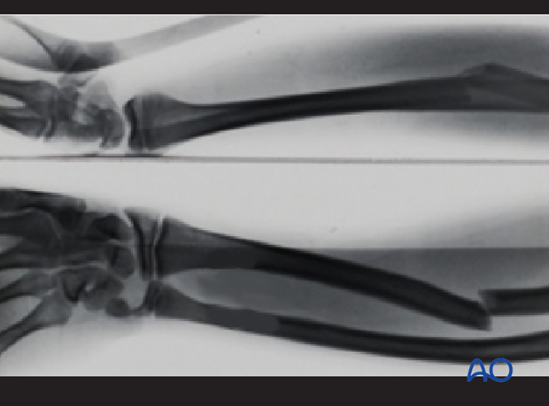
Transverse fracture of the radius and oblique fracture of the ulna, with both bones in apposition
