Assessment of rotation
1. Introduction
Confirm rotational alignment of the femur clinically and radiographically before finalizing fixation. This can be done by:
- Direct visualization of fracture site if open
- Fluoroscopy of fracture site
- Comparing internal and external rotation to the contralateral side
- Fluoroscopy of proximal femur (lesser trochanter profile and/or matching the cortices of both sides of the fracture)
2. Clinical assessment of rotation of the femur
Prior to patient preparation and draping, examine the opposite intact femur to determine the rotational range-of-motion.
If the patient is supine, demonstrate femoral rotation with the hip and knee each flexed to 90°.
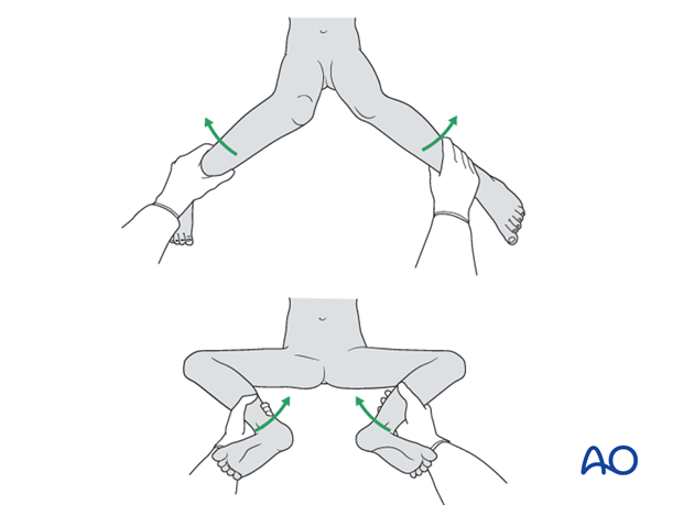
Internally and externally rotate the femur and record the maximum range.
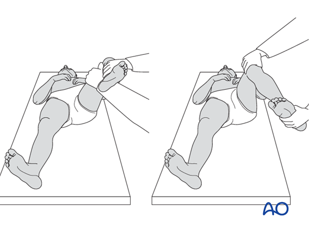
After reduction and (provisional) fixation of the fractured femur, examine internal and external rotation. They should be similar to the preoperative findings on the opposite side.
3. Fluoroscopy assessment of the lesser trochanter profile
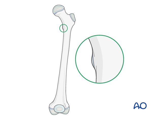
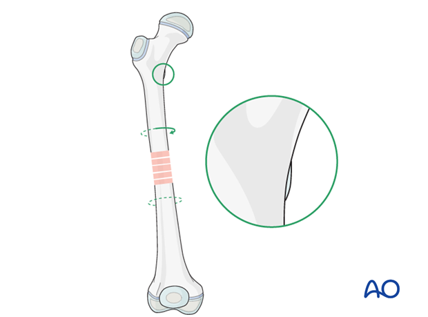
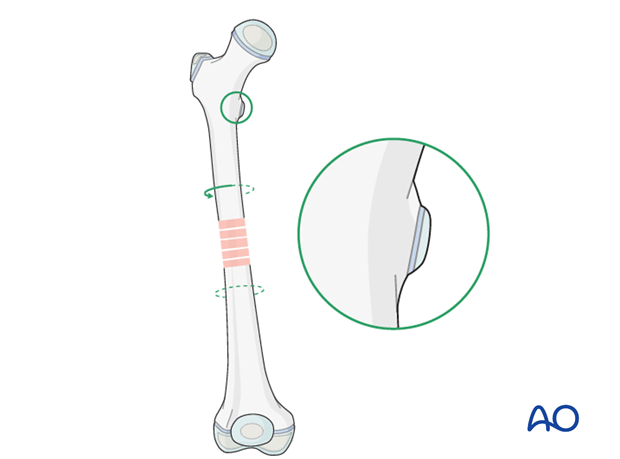
Compare the profile of the lesser trochanter on the intensifier image with that of the contralateral leg (lesser trochanter shape sign).
Before positioning the patient, store the profile of the lesser trochanter of the intact opposite side (patella facing anterior) in the image intensifier.
The illustration shows the lesser trochanter of the intact opposite side.
Malrotation
In cases of malrotation, the lesser trochanter is of different profile when compared to that of the contralateral leg.
Care should be taken to assess rotation with the patella facing directly forwards.
This illustration shows internal malrotation of the proximal fragment.
This illustration shows external malrotation of the proximal fragment.
Matching of the lesser trochanter shape
When rotational alignment is correct, the lesser trochanter profile will match that of the opposite side when the patella is facing forwards.

4. Fluoroscopy assessment of the cortical congruency across fracture site
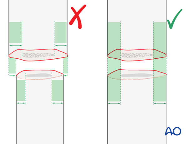
The appearance of the cortices across the fracture site is checked under image intensification.
Cortical thickness and shaft diameter should match.
